The temporal bones are a pair of bones located on either side of the skull, behind the ears. These bones have several important functions in the body.
First and foremost, the temporal bones serve as protective structures for the brain. They contain the auditory tubes, which allow for the passage of sound waves to the inner ear, and the auditory ossicles, which are small bones that transmit sound waves from the eardrum to the inner ear. The temporal bones also house the structures that are responsible for balance, including the vestibular system and the vestibulocochlear nerve.
In addition to their role in hearing and balance, the temporal bones also serve as attachment points for several muscles. These muscles, including the temporalis and masseter muscles, are responsible for chewing and facial expressions.
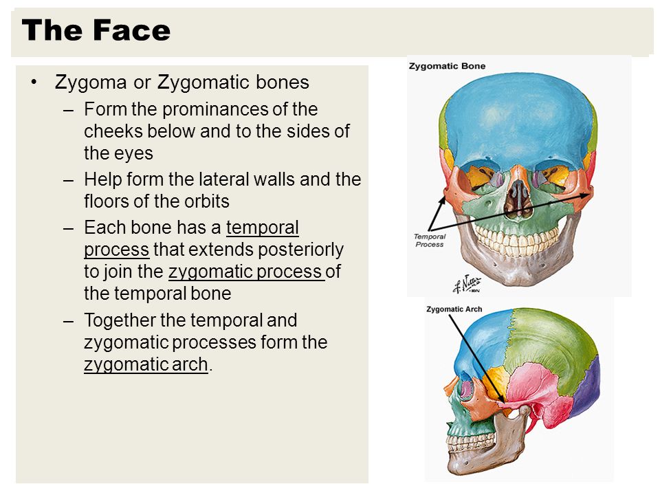
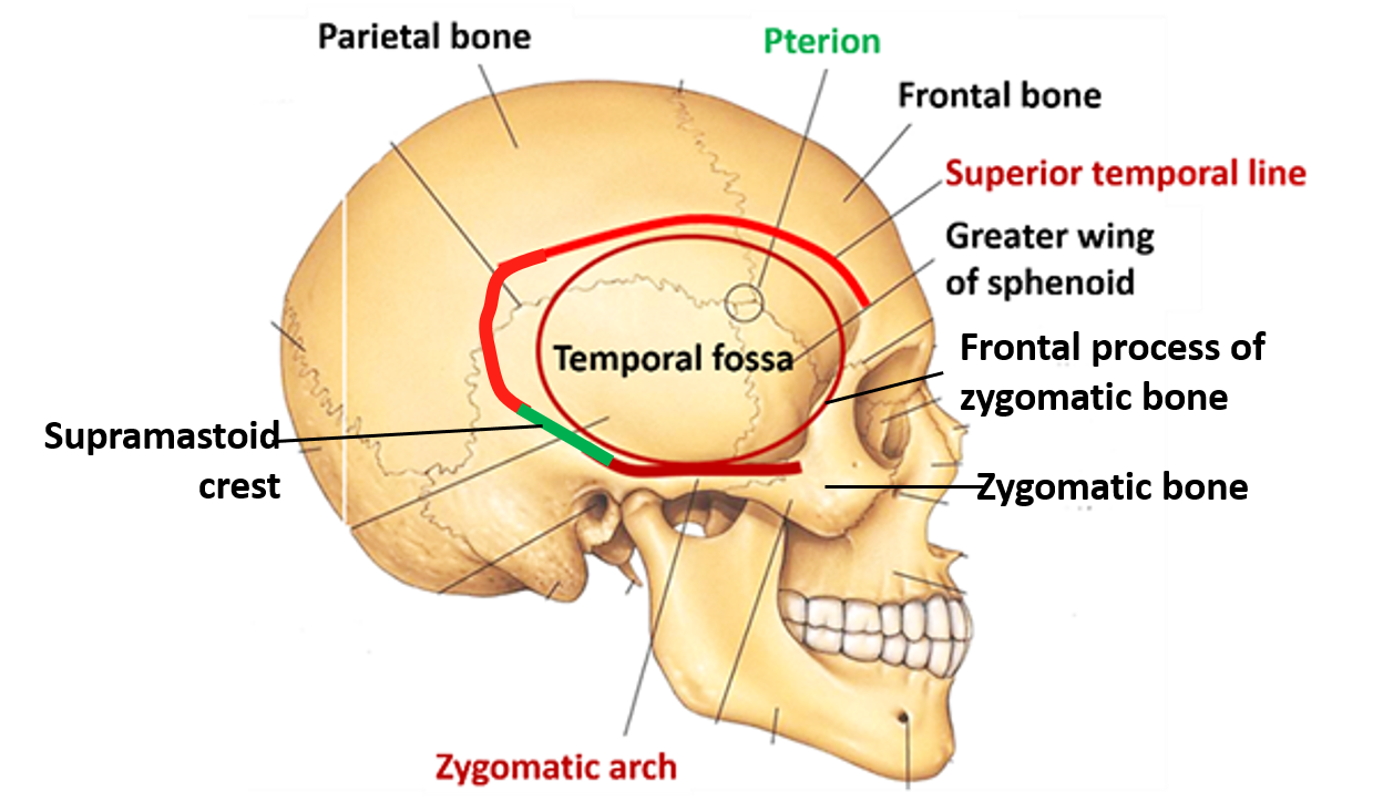
The temporal bones also contain the zygomatic arch, which is a bony arch that spans across the cheek and is important in the movement of the lower jaw. The zygomatic arch is formed by the zygomatic process of the temporal bone and the zygomatic process of the cheekbone.
Finally, the temporal bones contain several important blood vessels and nerves, including the internal carotid artery and the middle meningeal artery, which provide blood to the brain, and the trigeminal nerve, which is responsible for sensation in the face.
In summary, the temporal bones are essential for several important functions in the body, including hearing, balance, facial expression, and the supply of blood and nerves to the brain. They play a vital role in the overall function and health of the body.
Temporalis: Origin, insertion, innervation, function

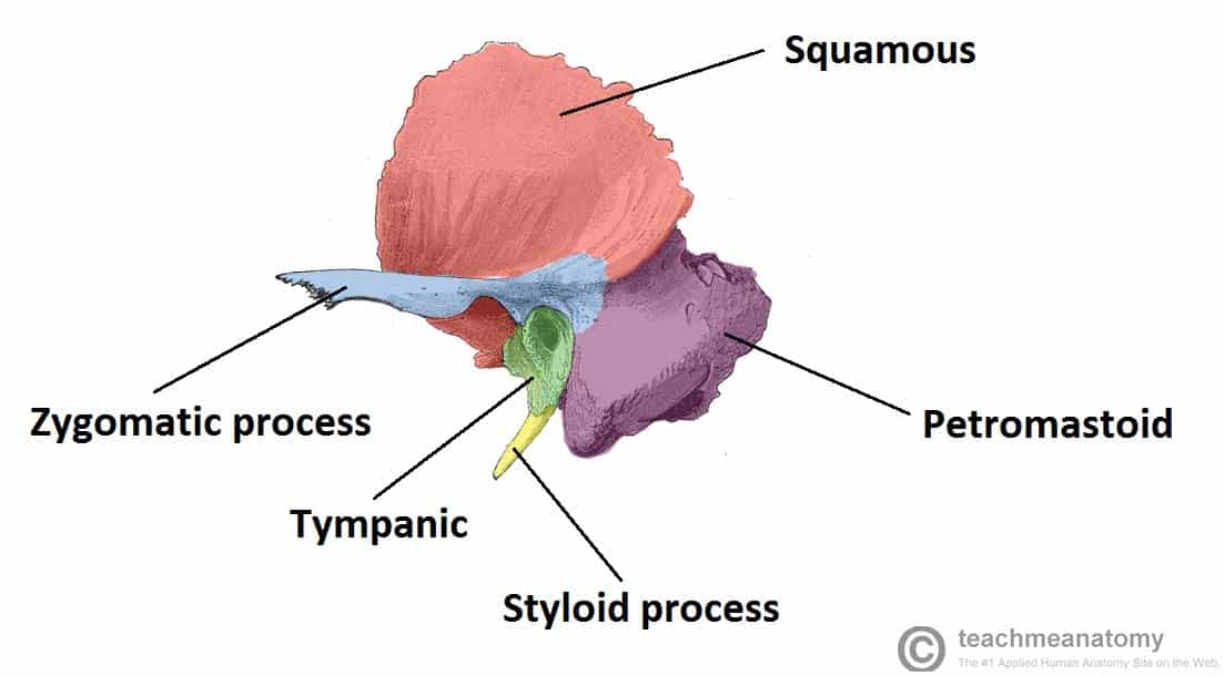
Ossification of the temporal bone, or bone development from cartilage, begins at 16 ta week, with the formation of the so-called temporal tympathetic rings. The commonest complication of this injury is the damage to the auriculotemporal nerves that run in close proximity to the joint. Figure 8A shows the relationship between the SCCs. The arrow is pointing to the zygomatic process The zygomatic bone, in case you're wondering, is shown in the following image. Hamid Djalilian and Harrison Lin , two head and neck cancer specialist Drs. The vestibulocochlear nerve terminates in the temporal bone. Simplify your learning and focus on the key aspects using Kenhub's Innervation The temporalis muscle receives its innervation from the anterior, middle and posterior deep temporal branches of the anterior trunk of the To expand your knowledge check out our other articles, videos, quizzes and labeled diagrams about the muscles of mastication.
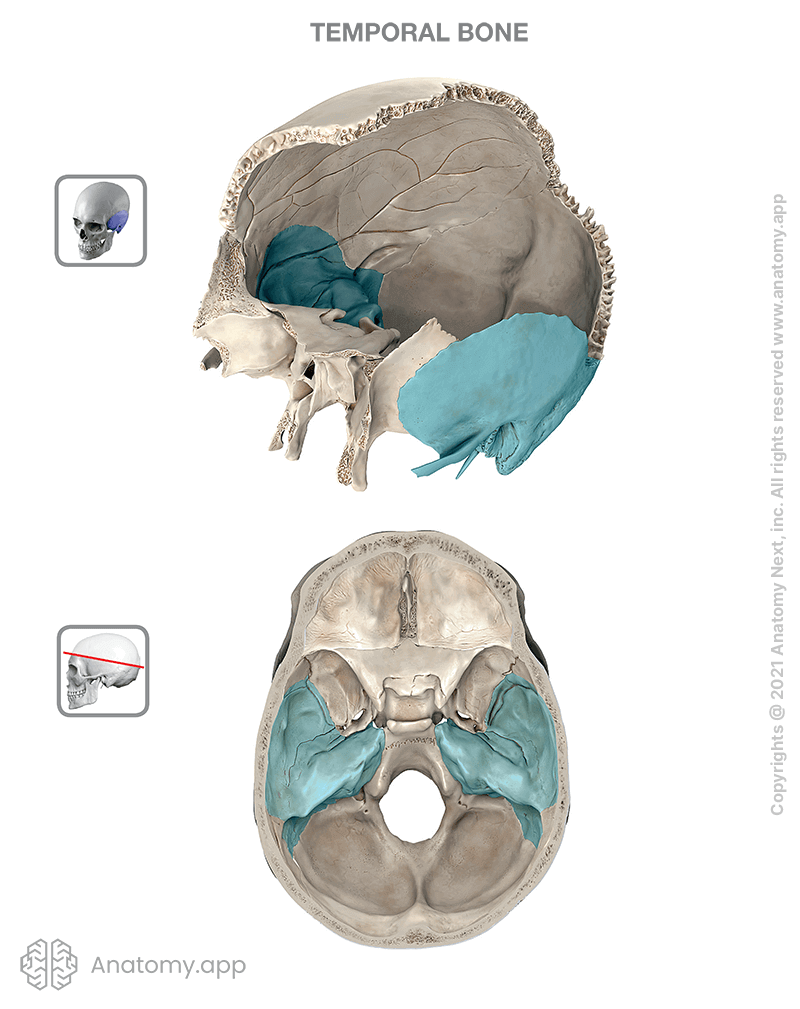
Temporal bone: Anatomical diagram, function, and injuries

We ensure an individualized plan for every patient and provide the information necessary for the patient to make an informed decision. Wernicke's Area This portion of the brain is very important for being able to process and understand the words of others. Next, carry the dissection from the zygomatic root towards the glenoid fossa of the temporalmandibular joint superiorly and the parotid inferiorly Figure 13A. If you feel your face, the zygomatic process helps to make up those lovely cheekbones you have! Edinburgh: Elsevier Churchill Livingstone. Below it articulates with the parietal bone. This protrusion is evaluable on physical examination, it can be felt behind the ear. What is the temporal bone? This can lead to a number of serious complications, including damage to hearing, vertigo, facial paralysis due to damage to the facial nerve , and bleeding in the ear as well as bone bruising.