How does a gamma camera work. Gamma Camera: Diagram, Isotopes, Scan & Applications 2022-10-10

How does a gamma camera work Rating: 4,5/10 1071 reviews

A gamma camera, also known as a scintillation camera or a gamma ray camera, is a medical imaging device that is used to produce images of the body for diagnostic and therapeutic purposes. It works by detecting gamma rays, which are a type of high-energy radiation emitted by certain types of radioactive substances.

The process begins when a small amount of a radioactive tracer, or isotope, is introduced into the body. This tracer is usually injected into the bloodstream, inhaled, or ingested, depending on the part of the body being imaged. The tracer accumulates in the cells and tissues of the body and emits gamma rays as it decays.

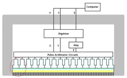
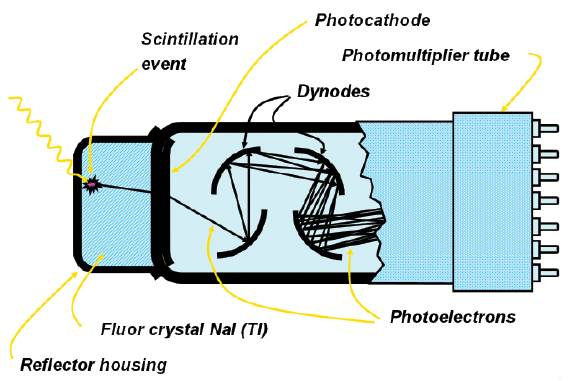
The gamma camera consists of a detector, which is a device that can detect and measure the energy of the gamma rays, and a computer that processes the data collected by the detector. The detector is typically made up of a crystal scintillator, which is a material that absorbs the energy of the gamma rays and emits light in proportion to the energy absorbed. This light is then detected by a photomultiplier tube, which converts the light into an electrical signal.

When the patient is placed in front of the gamma camera, the detector scans the body and detects the gamma rays emitted by the tracer. The computer processes this data and produces an image of the distribution of the tracer in the body. This allows doctors to visualize the function and structure of the organs and tissues being imaged and to detect any abnormalities.

There are several different types of gamma cameras, including single-headed and multi-headed cameras. Single-headed cameras are typically used for imaging smaller areas of the body, while multi-headed cameras are used for imaging larger areas or for performing more complex studies.

Gamma cameras have a number of advantages over other imaging modalities, such as X-rays and computed tomography (CT) scans. They are non-invasive and do not use ionizing radiation, which means that they do not pose the same risk of radiation exposure as other imaging modalities. In addition, they can provide functional information about the body, as they show how the organs and tissues are functioning, rather than just their structure.

In conclusion, a gamma camera is a medical imaging device that produces images of the body by detecting gamma rays emitted by a radioactive tracer. It is a non-invasive and safe way to visualize the function and structure of the body's organs and tissues and to detect abnormalities.

How does a gamma camera work Flashcards

how does a gamma camera work

Since gamma encoding redistributes tonal levels closer to how our eyes perceive them, fewer bits are needed to describe a given tonal range. The construction of a gamma camera is sometimes known as a compartmental radiation construction. Gamma rays are a radiation hazard for the entire body. This refers to the net influence of your video card and display device, so it may in fact be comprised of several gammas. Where is the collimator located on the gamma camera? What are gamma rays wavelength? In contrast to this, during " Positron Emission Tomography PET ", which nowadays as a so-called hybrid scanner is combined with CT or MRT, the detector is positioned in a ring shape tube-like around the patient. They differ in mass, energy and how deeply they penetrate people and objects. These are: Gas-Filled Detectors, Scintillators, and Solid State detectors.

Next

Understanding Gamma Correction

how does a gamma camera work

By measuring the intensity of radiation, the concentrations of medical tracer can be visualised, allowing diagnosis of the body processes and functions. Finally, in the fourth picture the display gamma over-compensates for the image gamma, resulting in an overall system gamma that curves downward thereby darkening the image. A higher display gamma results in a darker image with greater contrast. Can XRAY be scanned? The crystal gives a tiny flash of visible light every time a gamma photon hits it. When a gamma photon leaves the patient, it knocks an electron loose from an iodine atom in the crystal, and a faint flush of light is produce when the dislocated electron again finds a minimal energy state. More sophisticated gamma cameras have collimators that allow gamma photons from one point to go through several holes.


Next

Why is a collimator needed in a gamma camera?

how does a gamma camera work

Clever software and clever arrangement of the holes allows a brighter image to be reconstructed. These systems have been adapted for use in PET scan procedures. In that case, the file gamma may represent a combination of the encoding gamma and the camera's gamma. What are the pros and cons of gamma rays? How is a collimator used in nuclear medicine? If you experience any non-subsiding allergic reaction inform your doctor immediately. Because the window has a finite width, some scattered photons may still be accepted; e. Gamma cameras are used to construct an image of the distribution of radiopharmaceuticals spread out in the body of a patient.

Next

Gamma Camera

how does a gamma camera work

Although, the machine will not touch you, it may come very close to you during some scans. A gamma encoded image has to have "gamma correction" applied when it is viewed — which effectively converts it back into light from the original scene. Following the treadmill test, there is another waiting period followed by a second imaging scan. This test of tomographic resolution and uniformity should be performed every 6 months. These are radioisotopes combined with another molecule, such as glucose, which the body transports. How does a gamma detector work? While you are walking on the treadmill, a second small amount of Myoview is administered.

Next

Imaging in medicine: 6.3 How a gamma camera works

how does a gamma camera work

How much does a gamma camera cost? What rays come from space? Does sun emit gamma rays? The PMTs are arranged in a hexagonal grid, with the electrical pulse output of each being connected to a computer. The next step is where most people get confused, so take this part slowly. LCD monitors weren't so fortunate; ensuring an overall display gamma of 2. From the exterior to interior a camera is comprised of 1: head cover: usually made from glass. It works to a different principle to x-ray equipment, whereby an intensive x-ray beam penetrates the patient and illuminates the film behind it for a short while.

Next

What is gamma camera used for?

how does a gamma camera work

Depicts an image in the sRGB color space which encodes using a gamma of approx. WHY GAMMA IS USEFUL 1. This technique is known as Scintigraphy. What is Gamma in photography? Fortunately this second step the "display gamma" is automatically performed by your monitor and video card. It's a good idea to first have an How to interpret the charts. What crystals are used in gamma cameras? Note that because of the high counts present in such an acquisition, it is often necessary to acquire a high-count uniformity correction map ~100 Mcts or more in order to provide adequate uniformity correction for this study. Does PET use gamma camera? This is usually already set by the manufacturer's default settings, but can also be set during LCD Monitors.


Next

What is the science behind a gamma camera scan?

how does a gamma camera work

A tomographic version of the gamma camera is called SPECT, which yields slices through the body. The camera accumulates counts of gamma photons, which are detected by crystals in the camera. In PET the radionuclides used produce positrons. A radioactive source emits gamma ray photons in all directions. As soon as your reports are ready an SMS will be sent to the mobile number registered with Medanta. The services once availed cannot be cancelled except in cases where the User requests to cancel the service on the day of purchase in which case, full amount of service fees shall be refunded to the User provided that the Device has not been unboxed.

Next

Gamma Camera Imaging Technique

how does a gamma camera work

Detection of gamma radiation is one of the most important research tools in nuclear physics. However, the amount of radioactivity given in this procedure is strictly within the recommended national level. Do CT scans use gamma rays? A small amount of such medication is administered to patients and this produces a minute amount of radiation from the body that lasts for a limited time and can be picked up by the Gamma camera. Before we look at a patient being imaged and some of the images which can be obtained using this technique, we will look in a bit more detail at how a gamma camera works. What is a CT PET scan? SPECT Single-Photon Emission Computed Tomography is a diagnostic imaging technique used in nuclear medicine which studies PHYSIOLOGICAL FUNCTIONAL processes in the body. The gamma camera itself does not emit any radiation. What are the 4 types of radiation? Whenever gamma photon hits the crystal, it gives a tiny flash of visible light.

Next

How does a gamma camera work?

how does a gamma camera work

Doctors use PET-CT scans to provide more information about the cancer. What is the principle operating of gamma camera? The camera typically comprises of a flat and narrow table on which the patient must lie down. Technical Note on Camera Gamma. Which of the following detector is used for gamma ray detection? Gamma correction is sometimes specified in terms of the encoding gamma that it aims to compensate for — not the actual gamma that is applied. How does gamma camera detect radiopharmaceuticals? A higher gamma correction value might therefore brighten the image the opposite of a higher display gamma. The first picture far left gets brightened substantially because the image gamma — is uncorrected by the display gamma — , resulting in an overall system gamma — that curves upward. Gamma radiation, unlike alpha or beta, does not consist of any particles, instead consisting of a photon of energy being emitted from an unstable nucleus.

Next