Intrinsic conduction system definition. What is the intrinsic conduction system? 2022-11-03
Intrinsic conduction system definition Rating:
5,2/10
893
reviews
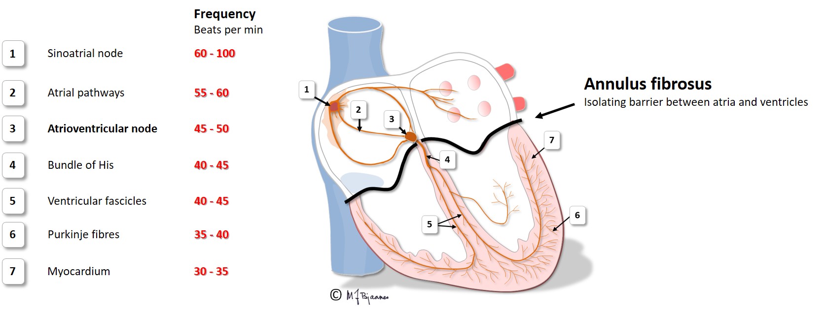
The intrinsic conduction system is a network of specialized cells found in the heart that regulate its contractions and coordinate the pumping of blood throughout the body. This system is made up of three main components: the sinoatrial (SA) node, the atrioventricular (AV) node, and the bundle of His.
The SA node, also known as the pacemaker of the heart, is located in the upper right chamber of the heart (the right atrium). It is responsible for generating and transmitting electrical signals that initiate the contraction of the heart muscle. The SA node has the highest intrinsic firing rate of any of the cells in the intrinsic conduction system, meaning it is able to generate electrical signals at a faster rate than any of the other cells.
The AV node is located in the lower right chamber of the heart (the right ventricle) and serves as a relay station for the electrical signals generated by the SA node. It receives the signals from the SA node and then transmits them to the bundle of His, which carries the signals to the rest of the heart.
The bundle of His is a bundle of specialized fibers that runs from the AV node down through the septum (the wall separating the left and right sides of the heart) and into the left ventricle. It carries the electrical signals generated by the SA node and AV node to the rest of the heart, allowing for coordinated contraction of the heart muscle.
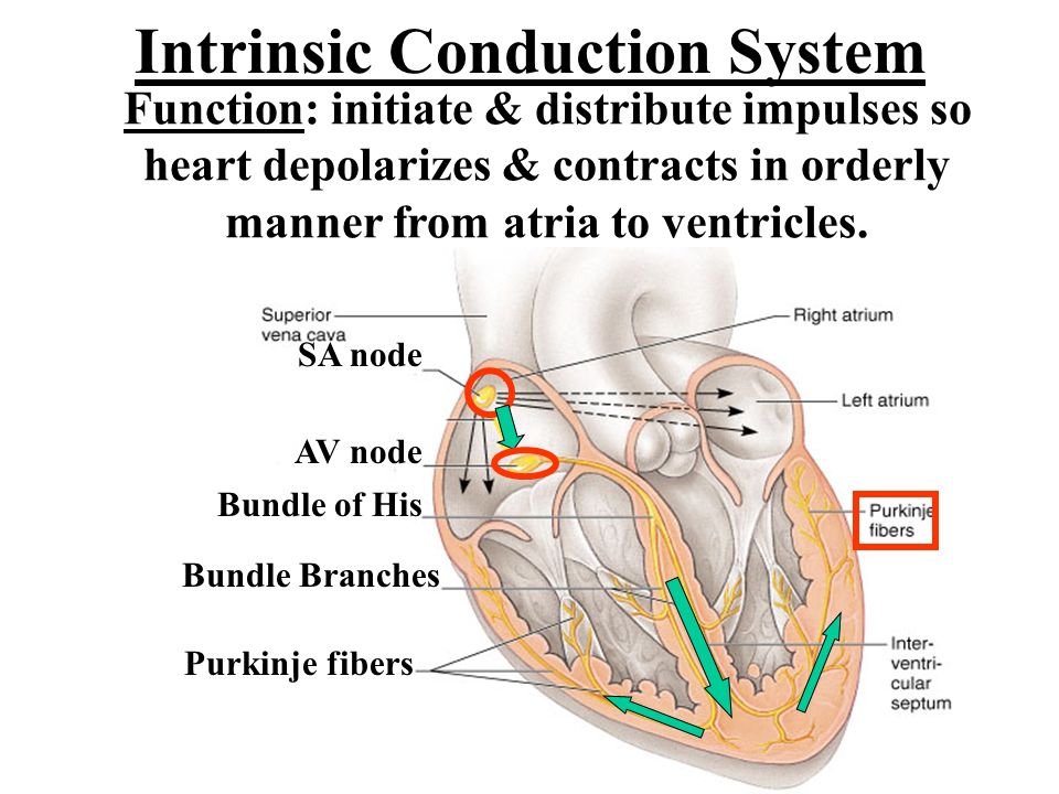
Together, the SA node, AV node, and bundle of His form the intrinsic conduction system, which is responsible for maintaining a regular heartbeat and coordinating the pumping of blood throughout the body. Disruptions in the function of this system can lead to abnormal heart rhythms, known as arrhythmias, which can be dangerous and even life-threatening.
Function of the Heart's Electrical System
You can wear these monitors for days or even weeks. The conduction system of the heart. Springer Publishing Company; 2020:3-8. This includes feeling like your heart is pounding, racing or skipping beats. So sushu -273 ° C, the semiconductor material into islator. The pressure in the ventricle continues to increase.
The SA node starts the sequence by causing the atrial muscles to contract. The cells that create these rhythmic impulses, setting the pace for blood pumping, are called pacemaker cells, and they directly control the heart rate. What are the elements of the intrinsic conduction system of the heart? Cardiac muscle cells can and do contract spontaneously and independently, even if all nervous connections are severed. Major Arteries of the Systemic Circulation The major branches of the aorta and the organs they serve are listed next in sequence from the heart. Distinct cells, each with one nucleus. The sinoatrial SA node is the pacemaker of the intrinsic conduction system of the heart. The valves of the heart secure a one-way blood flow through the heart and blood vessels.
What is the order of the intrinsic conduction system in order?
The slowing effect gives the ventricles enough time to expand and fill up with blood. Please visit our nursing test bank page for more NCLEX practice questions. At the end of systole, the ventricles relax, the semilunar valves snap shut, and for a moment the ventricles are completely closed chambers; the intraventricular pressure drops and the AV valves are forced open; the ventricles again begin refilling rapidly with blood, completing the cycle. As the ventricle contracts, blood leaves the heart through the pulmonic valve, into the pulmonary artery and to the lungs where it is oxygenated. It occurs at the beginning of ventricular systole and results from closure of the AV valves. Its broad posterosuperior aspect, or base, from which the great vessels of the body emerge, points toward the right shoulder and lies beneath the second rib.
It is considered as the bluntly rounded portion of the heart A. The first step of cardiac conduction is impulse generation. The intrinsic conduction system of the heart is comprised of several specialized subpopulations of cells that either spontaneously generate electrical activity pacemaker cells , or preferentially conduct this excitation throughout the four chambers of the heart in a coordinated fashion. What is the function of the intrinsic conduction system of the heart quizlet? They occur at the Z-line of the sarcomere and can be visualized easily when observing a longitudinal section of the tissue. Anatomy of the Heart The cardiovascular system can be compared to a muscular pump equipped with one-way valves and a system of large and small plumbing tubes within which the blood travels. What are the five steps in the conduction system? K + channels to close quickly, decreasing the permeability of the cell membrane to K +. .
intrinsic conduction system of the heart Flashcards
Which of these statements correctly applies to intrinsic regulation of the heart? Extrinsic Conduction Electronic components such as diodes and transistors made of semiconductor materials. It then passes rapidly through the AV bundle. The branches carry the electrical cascade to the outer areas of the heart, especially the ventricles. The myocardium is drained by several cardiac veins, which empty into an enlarged vessel on the posterior of the heart called the coronary sinus. Care What can I do to help my cardiac conduction system? The myocardium consists of thick bundles of cardiac muscle twisted and whirled into ringlike arrangements and it is the layer that actually contracts. This includes your heart, breathing, digestion and more.
Cardiovascular System Anatomy and Physiology: Study Guide for Nurses
While all cells in your heart can conduct electricity, the cells in this system conduct it at very specific speeds. The pulmonary veins empty oxygen-rich blood from the lungs into the left atrium. This includes pacemakers or implantable cardioverter defibrillators. This system causes heart muscle depolarization in only one direction- from the atria to the ventricles; it enforces a contraction rate of approximately 75 beats per minute on the heart, thus the heart beats as a coordinated unit. As the ventricle contracts, blood leaves the heart through the aortic valve, into the aorta and to the body. Gap junctions ADVERTISEMENTS D: Gap junctions are a specialized intercellular connection between a multitude of animal cell-types. The right AV valve, the tricuspid valve, has three flaps.
What is the pacemaker of the intrinsic conduction system?
What is the electrical conduction pathway of the heart? More round when relaxed, oval-shaped when flexed. The subclavian vein receives venous blood from the arm through the axillary vein and from the skin and muscles of the head through the external jugular vein. P-Q or P-R interval C. The heart conducts electricity in a specific way as described above, and its function works best when it maintains this sequence. What are pacemaker cells? Cardiac Output Cardiac output is the amount of blood pumped out by each side of the heart in one minute. The radial and ulnar veins are deep veins draining the forearm; they unite to form the deep brachial vein, which drains the arm and empties into the axillary vein in the axillary region. The vertebral vein drains the posterior part of the head.
What is the important function of the intrinsic conduction system?
As the atrium contracts, blood flows from the right atrium to the right ventricle through the open tricuspid valve. As a result, the semilunar are forced open and blood is ejected into the pulmonary trunk and aorta. The basilic and cephalic veins are joined at the anterior aspect of the elbow by the median cubital vein, often chosen as the site for blood removal for the purpose of blood testing. To do this, it relies on a specialized network of cells called the cardiac conduction system. The hepatic veins drain the liver. Atrioventricular or AV valves are located between the atrial and ventricular chambers on each side, and they prevent backflow into the atria when the ventricles contract. The main parts of the system are the SA node, AV node, bundle of HIS, bundle branches, and Purkinje fibers.