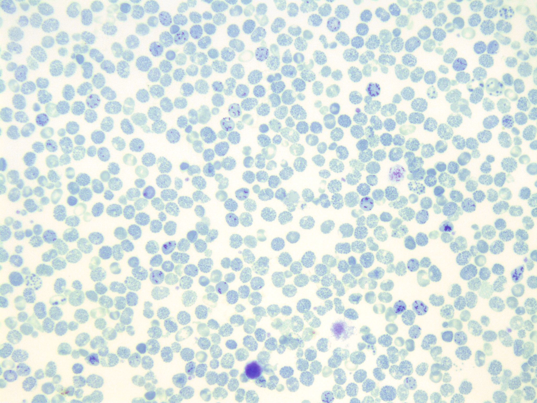
HBH inclusion bodies are small, spherical structures that can be found within cells. They are made up of a protein called HBH, which stands for "host cell protein." HBH inclusion bodies are often found in cells that have been infected with a virus, as the virus will often produce HBH as a way to protect itself from the host cell's immune system.
The presence of HBH inclusion bodies is often used as a diagnostic tool to detect viral infections. For example, if a person is experiencing symptoms of a viral infection, a sample of their cells may be examined under a microscope to see if HBH inclusion bodies are present. If they are, it is likely that the person has a viral infection.
HBH inclusion bodies can also be found in cells that have been genetically modified to produce large amounts of a particular protein. In these cases, the inclusion bodies are formed as a way for the cell to store and protect the protein.
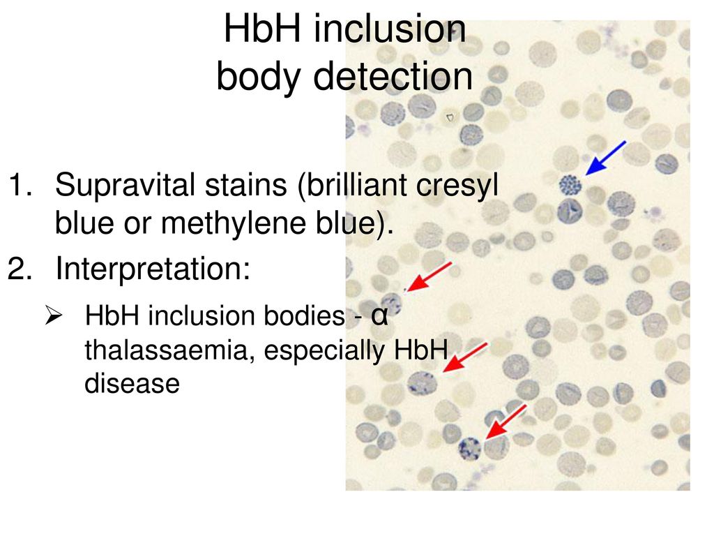
There are several ways in which HBH inclusion bodies can be visualized and analyzed. One common method is to stain the cells with a fluorescent dye that specifically binds to HBH, allowing the inclusion bodies to be seen under a microscope. Another method is to use electron microscopy, which allows for a more detailed view of the inclusion bodies.
Despite their potential usefulness as a diagnostic tool, HBH inclusion bodies can also be a problem for scientists and researchers working with genetically modified cells. This is because the presence of inclusion bodies can indicate that the cell is not functioning properly, and that the protein being produced is not being properly folded and transported within the cell.
In conclusion, HBH inclusion bodies are small, spherical structures that are made up of the protein HBH. They can be found in cells that have been infected with a virus or genetically modified to produce large amounts of a particular protein. While they can be useful as a diagnostic tool, their presence can also indicate problems with the function of the cell.
Alpha
Retrieved 22 September 2016. Hemoglobin A2 HbA2 makes 1% to 3% of adult hemoglobin. Novel large deletions in the human alpha-globin gene cluster: Clarifying the HS-40 long-range regulatory role in the native chromosome environment. However, because this disorder is underdiagnosed it is difficult to determine its true frequency in the general population. The molecular tests are not widely available in most of the laboratories and also not cost effective. The specificity and simplicity of the immunocytochemical test make it the test of choice in screening for alpha thalassaemia. Alpha thalassemia, a condition where there is a defect in the production of the oxygen-carrying pigments of red blood cells hemoglobin , is not seen in every case.
Figure adapted with permission from Cambridge University Press Blood. Hemoglobin F HbF is comprised of two alpha and two gamma chains. Figure adapted with permission from Cambridge University Press Blood. The α subunit lacks helix D. Louis, Missouri: Elsevier Inc.
Hemoglobin H disease: not necessarily a benign disorder
Introduction Some researchers have suggested the name XLID-hypotonic face syndrome be used to designate several disorders formerly considered separate entities including ATR-X syndrome, Carpenter-Waziri syndrome, Chudley-Lowry syndrome, Holmes-Gang syndrome and X-linked intellectual disability-arch fingerprints-hypotonia syndrome. It can bring about growth retardation during childhood and iron overload in adults regardless of previous transfusion history, leading to hepatic, cardiac, and endocrine dysfunction. The condition is called thalassemia minor. In non-deletion, the severity of clinical expression is also affected depending on whether the mutation blocks the production of the remaining normal alpha chains partially or fully. Retrieved 15 September 2016. However, HbH is a variable finding in ATR-X syndrome and failure to detect HbH inclusion bodies does not rule out ATR-X syndrome.
These people usually have moderate anemia and marked microcytosis and hypochromia. ATR-X syndrome affects males. Gibbons RJ, McDowell TL, Raman S, et al. This is characterised by microcytosis, and occasional target cells. No diagnosis was reached for the remaining patient. Growth and development were normal in all of our patients. Am J Med Genet.
Hemoglobin H disease causes, symptoms, diagnosis & treatment
Beta thalassemia major is caused by a homozygous mutation beta-zero thalassemia of the beta-globin gene, resulting in the total absence of beta chains. HbH disease is an uncommon clinical phenotype of α-thalassemia with variable clinical presentation. There is a wide spectrum of genotypes and phenotypic presentations. Many affected individuals have abnormalities of the genitourinary tract including failure of the testes to descend into the scrotum cryptorchidism , unusual placement of the urinary opening meatus on the underside of the penis hypospadias , an abnormal fold of skin extending around the base of the penis shawl scrotum , and underdevelopment of the scrotum. CONCLUSION: We emphasize a systematic screening of hemograms and peripheral smears along with a simple and cost-effective screening test for golf ball inclusions to diagnose HbH disease that often poses a clinical dilemma due to its variable clinical presentation. Population genetics Hb H disease is found in many parts of the world, including Southeast Asian, Middle Eastern, and Mediterranean populations. Patients would then have the typical presentation of hemolytic anemia, indirect hyperbilirubinemia, elevated LDH, decreased haptoglobin, splenomegaly, hepatomegaly, jaundice, variable bony changes, premature biliary tract disease, leg ulcers, and eventually iron overload if multiple transfusions are required without adequate iron chelation.
Low hemoglobin with low red blood cell count and low hematocrit indicates anemia. HbH disease should be considered in infants or children with mild-to-moderate microcytic hypochromic hemolytic anemia and hepatosplenomegaly. Picketts DJ, Higgs DR, Bachoo S, et al. Hb biochemical analysis reveals the presence of HbH 5-30%. HbH patients are at risk of clinical manifestations with oxidative damage. Significant anemia might occur during infections, fever, hypersplenism, or pregnancy that may necessitate the need for blood transfusions.
Comparison of haemoglobin H inclusion bodies with embryonic zeta globin in screening for alpha thalassaemia.
ATR-X syndrome is characterized by intellectual disability, characteristic facial features, abnormalities of the genitourinary tract, and alpha thalassemia. Thalassemia is the name of a group of genetic blood disorders caused by decreased synthesis of alpha or beta chains of hemoglobin Hb. New Jersey: Pearson; 2015. Utility of screening golf ball inclusions in hemoglobin H disease and its clinico-hematological profile. What does Hemoglobin H trace mean? X-linked intellectual disability update 2017.
Deferasirox reduces iron overload significantly in nontransfusion-dependent thalassemia: 1-year results from a prospective, randomized, double-blind, placebo-controlled study. This causes significantly impaired alpha globin production. Growth deficiency occurs after birth postnatally , but may not become apparent until adolescence. Keywords:Alpha-thalassemia,Golf ball inclusions, Hemoglobin H disease, Hemoglobinopathy How to cite this article: Chandra D, Tyagi S, Deka R, Chauhan R, Singh J, Seth T, Pati HP, Saxena R. The name ATR-X syndrome is the most widely-recognized term for this disorder.