Describe the structure of the ear. Hearing and the Structure of the Ear 2022-10-13

Describe the structure of the ear Rating: 9,1/10 1354 reviews

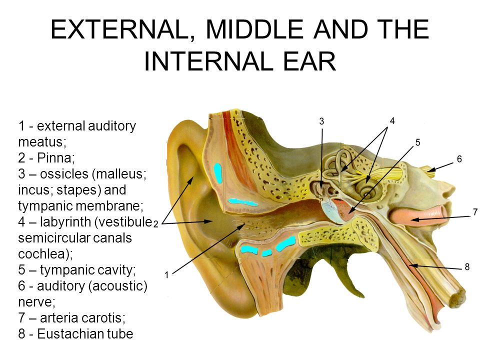
The ear is a complex organ that plays a vital role in our ability to hear and maintain balance. It is divided into three main parts: the outer ear, the middle ear, and the inner ear.

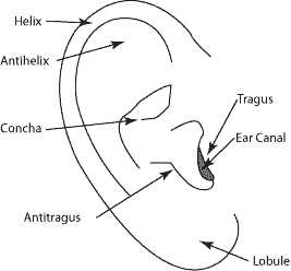
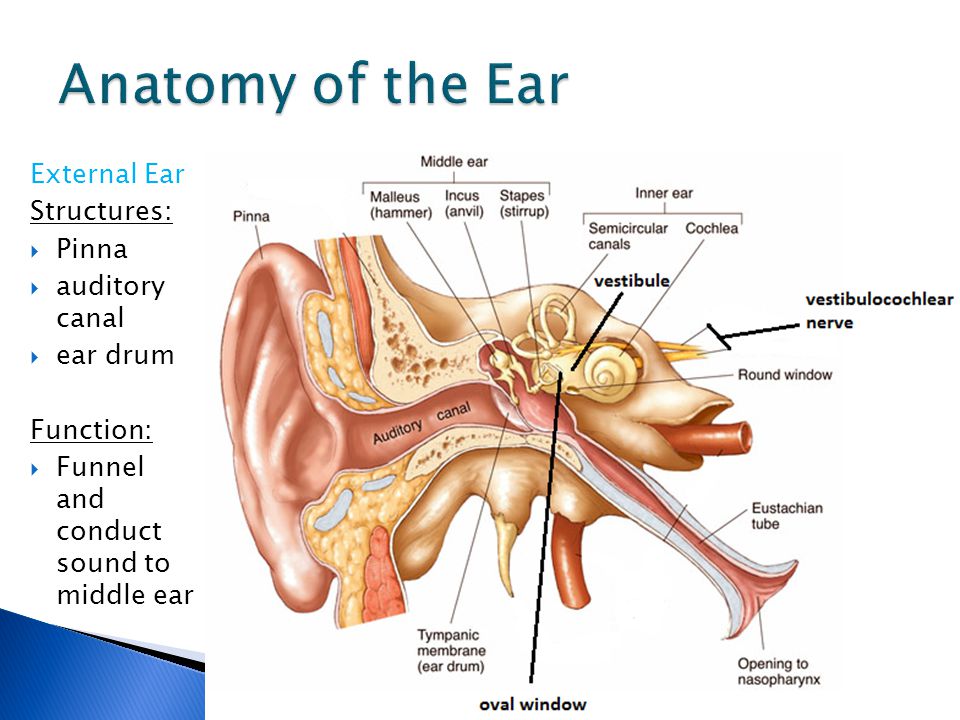
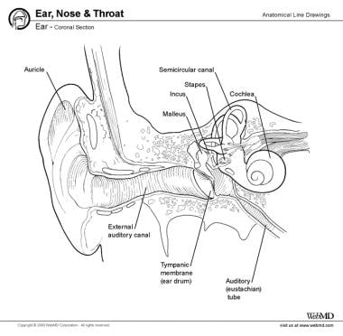
The outer ear consists of the visible part of the ear, called the pinna, and the ear canal. The pinna is the fleshy, visible part of the ear that is made up of cartilage and skin. It is responsible for capturing sound waves and directing them into the ear canal. The ear canal is a narrow, tubular structure that leads from the pinna to the eardrum. It is lined with thin, sensitive skin and helps to protect the inner ear from foreign objects and debris.

The middle ear is a small, air-filled chamber that is located behind the eardrum. It contains three small bones called the ossicles, which are the smallest bones in the human body. These bones are the malleus, incus, and stapes, also known as the hammer, anvil, and stirrup. The ossicles are connected in a chain and are responsible for transmitting sound waves from the eardrum to the inner ear. The middle ear also contains the Eustachian tube, which helps to maintain equal pressure on both sides of the eardrum.

The inner ear is a complex system of fluid-filled tubes and sacs that are responsible for converting sound waves into electrical signals that can be interpreted by the brain. It contains the cochlea, which is a snail-shaped structure that is responsible for converting sound waves into electrical signals, and the vestibular system, which is responsible for maintaining balance and detecting movement. The vestibular system consists of the vestibular nerve, the semicircular canals, and the vestibular sacs.

In conclusion, the ear is a complex organ that is made up of three main parts: the outer ear, the middle ear, and the inner ear. Each part plays a vital role in our ability to hear and maintain balance. The outer ear captures sound waves and directs them into the ear canal, the middle ear contains the ossicles and the Eustachian tube, and the inner ear contains the cochlea and the vestibular system. Together, these parts work to allow us to hear and maintain balance in our everyday lives.

The Human Ear

describe the structure of the ear

If the movement of fluid is great enough, the hairs are deflected and very small channels open up some­where near the tips of the hairs. Because of the natural curvature of the external acoustic meatus, the physician needs to pull the helix of the auricle posterosuperiorly, in order to align the meatus in a straight direction. The tectorial membrane overhangs the sensory hair in the scala media. Middle Ear: It includes the following: i The tympanic cavity, filled with air is connected with the nasopharynx through the Eustachian tube auditory tube , which serves to equalize the air pressure in the tym­panic cavity with that on the outside. Between the epithelial layers is connective tissue composed of collagen, elastic fibers, and fibroblasts. The doctor will likely use a lighted instrument an otoscope to look at the ears, throat and nasal passage.

Next

Human Ear: Structure and Functions (With Diagram)

describe the structure of the ear

Let's explore how the ear is broken down, its major parts, what their roles are in hearing and equilibrium, and understand how noises are processed and transmitted through the ear to help you to able to hear! The movements of the tectorial membrane stimulate the hair cells which then generate electrical impulses that transmit through the cochlear nerve to the Vasculature of the internal ear The bony labyrinth is vascularized by the anterior tympanic artery a branch of the maxillary artery , stylomastoid artery a branch of the posterior auricular artery , and the petrosal artery a branch of the middle meningeal artery. Kenhub does not provide medical advice. The central part of the tympanic membrane is called the umbo. The first canal directs the signals conducted into the liquid to the tip and back via a second canal. Stapes is also the smallest bone in the body.

Next

Briefly describe the structure of the ear.

describe the structure of the ear

An outer ear infection is usually considered chronic if signs and symptoms persist for more than three months. The three main structures of the ear are the outer ear, middle ear and inner ear. The hammer, anvil, and stapes are three bones inside the middle ear that sound passes through to reach the round window. Resting on the basilar membrane is the spiral organ or organ of Corti. The roots of the hairs produce oils and these mix with the secretions from nearby sweat-like glands to form the basis of wax.

Next

Ear anatomy: Parts and functions

describe the structure of the ear

They also have wax glands. It passes through the modiolus and the base of the cochlea. Internal Ear: There is a body cavity on each side enclosed in the hard periotic bone which contains the perilymph. One edge of this is attached to the bony core at the centre of the cochlea the modiolus ; the other is loosely attached to the organ of Corti outside the outermost outer hair cell. It divides both the outer ear and middle ear. This conflict between the two systems systems stimulates the area postrema the emetic center in the brain and presents as the nausea or even vomiting.

Next

The Structure of the ear

describe the structure of the ear

More medially, we find the oval window and round window, which separate the middle ear from the inner ear. Protect your ears from unnecessary damage by wearing protective earplugs or earmuffs in your workplace or during recreational activities if loud noise is present. SOUND AND HOW THE EAR WORKS Sound Sound travels as small waves of pressure through the air at a speed of about 343 metres per second 740 miles per hour. Pitches or the specific sound frequencies stimulate specific individual hair cells in the inner ear. Within the ampulla are special sensory cells called epithelia and hair cells underneath a substance called gelatinous copula.


Next

The Ear: Anatomy, Function, and Treatment

describe the structure of the ear

The following table will give you an idea of the various sound decibels that help human beings perceive different sounds. Inner Ear: Labyrinth is the scientific name of the inner ear. This means that the membranous labyrinth is suspended in the perilymph of the bony labyrinth. The first region is called the outer ear, which consists of the visible portion that you see when looking at yourself in the mirror. How the Human Ear Functions You can see in the diagram that the external ear captures the traveling sound waves. Acute otitis media occurs rapidly, is painful, and may cause fever.

Next

How the Ear Works

describe the structure of the ear

It is the smallest bone in the entire body. On the basilar membrane inside this duct is the spiral organ, which is the organ of hearing. These vibrations, which are transmitted through the fluid cause tiny hair cells in the fluid inside the cochlea to vibrate and stimulate the corti, thus generating nerve impulses that then travel to the brain. They pass through the external auditory meatus to the tympanic membrane which is caused to vibrate. Since vertigo is a symptom, and not a disease, its treatment depends on discovering its cause and treating it.

Next

Human Ear: Structure and Anatomy

describe the structure of the ear

It is mostly absorbed in the young kids within the age of 7 years old. The cause of balance problems is usually related to the specific sign or symptom. The three tiny bones forming the ossicles are malleus, incus and stapes. Even a cotton swab meant for cleaning earwax should not be put inside your ear canal because it can puncture your eardrum. Internally it consists of three fluid filled chambers or canals, the upper scala vestibuli, lower scala tympani, and the middle scala media cochlear duct. By contrast, 90% of the motor neurons in the cochlear nerve synapse with outer hair cells. It extends from the modiolus and attaches to the lateral wall of the cochlear duct.

Next

Structure of Ear: Anatomy and Physiology

describe the structure of the ear

Cell bodies of the sensory neurons are located in the spiral ganglion. The tympanic nerve and caroticotympanic nerves of the internal carotid plexus form the tympanic plexus in the mucous membrane that covers the promontory on the labyrinthine wall. The conductive hearing loss slowly progresses in early adulthood. The brain not only recognises the sound but also judges the direction, loudness and pitch of the sound. Managing pain Your doctor will advise you on treatments to lessen pain from an ear infection. It is composed of a group of interconnected canals and sacs.

Next