3 coats of the eye. What are the three layers of the human eye? 2022-10-13
3 coats of the eye Rating:
4,5/10
1138
reviews
The human eye is an incredibly complex and sophisticated organ, and it is made up of several layers or coats. These coats work together to protect and nourish the eye, and they also play a key role in helping us see the world around us. In this essay, we will explore the three main coats of the eye and how they function.
The first coat of the eye is called the sclera. This is the outermost layer and it is made up of a tough, white, fibrous tissue. The sclera is responsible for protecting the eye and helping it maintain its shape. It also helps to anchor the eye muscles, which allow us to move our eyes in different directions.
The second coat of the eye is called the choroid. This layer is located just beneath the sclera and it is made up of a thin, vascular layer of tissue. The choroid is responsible for providing oxygen and nutrients to the retina, which is the light-sensitive layer of cells at the back of the eye. It also helps to regulate the temperature of the eye and prevent it from overheating.
The third coat of the eye is called the retina. As mentioned earlier, the retina is a light-sensitive layer of cells that is located at the back of the eye. It is responsible for converting light into electrical signals that are sent to the brain via the optic nerve. The retina is made up of several different types of cells, including rods and cones. Rods are responsible for detecting light and dark, while cones are responsible for detecting color. Together, these cells help us see the world around us in all its detail and color.
In conclusion, the three coats of the eye – the sclera, choroid, and retina – work together to protect and nourish the eye, and to help us see the world around us. Each coat plays a vital role in the overall functioning of the eye, and without any one of these layers, our vision would be impaired.
What are the 3 coats of the eye?
When light is focused on this spot, visual acuity is as good as it gets. It is a transparent membrane that covers the front of the eye. This website includes study notes, research papers, essays, articles and other allied information submitted by visitors like YOU. You can see this reflex in action when you shine a low-intensity flashlight into a friend's eyes. At the level of the collarette of iris, the radial arteries anastomose with each other and form the minor arterial circle of iris. Humor If we look at the basic structure of the eyeball, we see that it's somewhat like a fluid-filled balloon that's covered by three coats. Deterioration of special light sensitive cells in the retina.
The mydriasis is a result of dilator pupillae muscle contraction. Iris The iris is a contractile, heavily pigmented, circular diaphragm that is analogous to the diaphragm of a camera. The cornea focuses light as it enters the eyes. Fovea Centralis The macula. Â Lens Synonyms: Crystalline lens, Lens crystallina The lens is a circular biconvex structure found anterior to the vitreous body and posterior to the iris.
The latter value decreases slowly with age; older people's eyes sometimes dilate to not more than 5—6mm in the dark, and may be as small as 1mm in the light. The refraction of light occurs in the center of the cornea, where its refractive power is significantly higher than that of the atmospheric air. The eyeball sits within the bony orbit of the skull. What part of the eye contains rods and cones? It's a small depression in the retina that contains the highest concentration of cones. This change in size regulates how much light is allowed to enter your eyes.
Eye Anatomy: 16 Parts of the Eye & Their Functions
The ciliary body contains smooth muscle that controls the shape of the lens. Rods and cones are photoreceptors in the retina. The cornea is described in the text above, so here we will focus on the lens, vitreous body and aqueous humor. Similarly, a deep cut or tear of the cornea, called corneal laceration, may occur if sharp objects such as fingernails or the corner of a page of a book comes in contact with the cornea. In fact, 70% of the eye's focusing power comes from the cornea and 30% from the lens. Glaucoma may develop if a person's aqueous fluid does not drain adequately.
The episclera is a thin layer of tissue that lies on top of the sclera. Several suspected causal factors in our environment have been studied so far. An injury of the iris can squeeze the iridocorneal angle and obstruct the aqueous humor outflow, which leads to a condition called closed-angle glaucoma. These drains are found at the base of the iris. Eyelashes protect the eye by catching dust and debris from entering the eye. Attaches to the top of the eye and moves the eye upwards.
The eye is made up of three coats. Which are they?
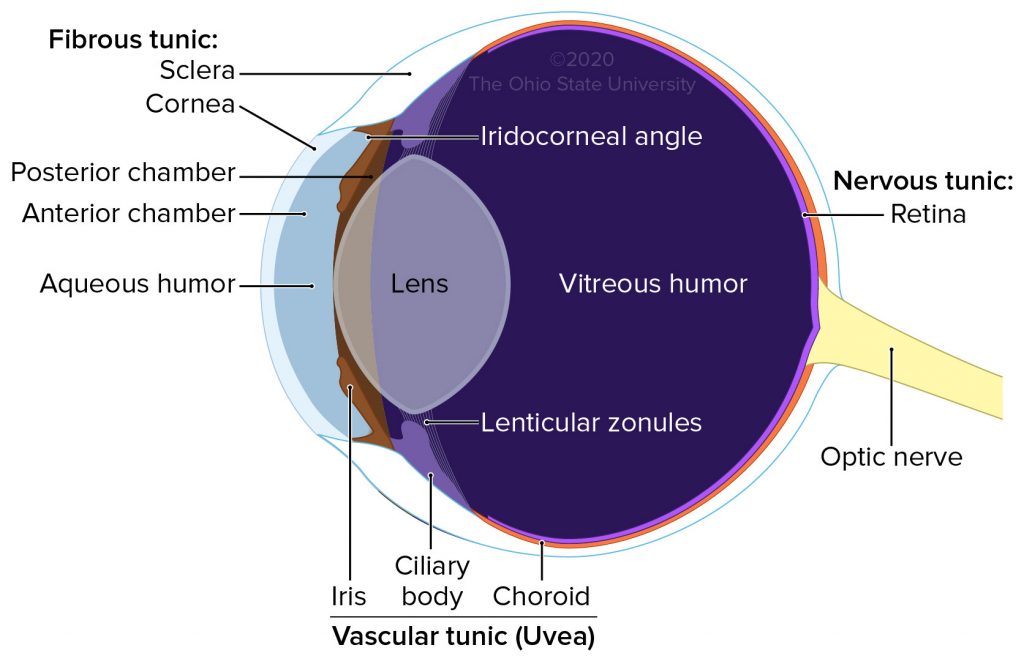
If this drainage canal gets blocked, pressure within the eye builds, leading to a condition known as glaucoma. If this drainage canal gets blocked, pressure within the eye builds, leading to a condition known as glaucoma. Physiological Basis of Aging and Geriatrics, Fourth Edition. Journal of Occupational Medicine. The eye is made up of three layers: the outer layer called the fibrous tunic, which consists of the sclera and the cornea; the middle layer responsible for nourishment, called the vascular tunic, which consists of the iris, the choroid, and the ciliary body; and the inner layer of photoreceptors and neurons called the … The eye is made up of three coats, or layers, enclosing various anatomical structures. The eyeball is generally less tall than it is wide. The most abundant supporting cells are the Müller cells, which are dispersed throughout the entire neural retina.
Pupils The pupil is seen as a black dot in the center of the iris. The fluid inside the eyeball is referred to as the humor. Review of Ophthalmology: Expert Consult — Online and Print. The prefix 'photo' means 'light,' and the suffix 'receptors' can be thought of as 'responders. What germ layer does the brain come from? The movements of the eye are controlled by six muscles attached to each eye, and allow the eye to elevate, depress, converge, diverge and roll. Light comes into it and bends evenly, which gives you a clear view. Usually, the anterior chamber is tinged red but blood soon accumulates in this chamber and vision becomes impaired.
Representative cases are presented at and Clinical diagnosis Besides thorough fundoscopy, which ultimately establishes the diagnosis, several clinical tests may be utilized. Caretta D; Tonali, P; Albanese, A. It is composed of light sensitive cells known as rods and cones. When the focus shifts to close objects, the ciliary muscles contract resulting in the relaxation of the suspensory ligament of lens. This is called the consensual light reflex and it occurs mainly because of the partial decussation of the optic nerve and optic tracts at the Hyphemia Hyphema or hyphemia is a condition in which there is hemorrhage within the anterior chamber of the eyeball.
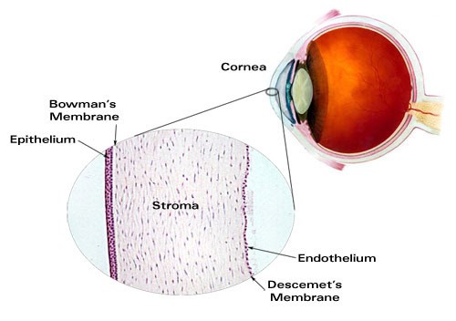
It contains vessels that carry blood through the eye to nourish it. Aqueous Humor Aqueous humor is a fluid substance that fills the eye. Clinical anatomy of the eye 2nd ed. The ciliary body and its muscles surround the lens to adjust its shape. The cones allow you to see colors. This is in fact the basement membrane of the underlying corneal endothelium and it consists of the collagen fibers.
%3A+1-+Fibrous+tunic%3A+Cornea.+Sclera..jpg)