Virtual labs are a valuable tool for educators, students, and researchers. They allow us to explore complex scientific concepts and processes without the need for expensive equipment or risky experimentation. One such virtual lab is the Cell Cycle and Cancer virtual lab, which allows users to explore the mechanisms behind cell division and how it can go wrong in the development of cancer.
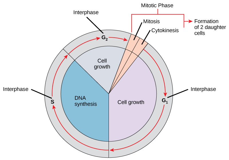
The cell cycle is the process by which cells grow and divide. It is made up of two main stages: interphase and mitosis. Interphase is the period of cell growth and DNA synthesis, during which the cell prepares for division. Mitosis is the actual process of cell division, during which the cell's nucleus and cytoplasm are divided into two identical daughter cells.
Cancer is a disease that is characterized by the uncontrolled growth and division of cells. It occurs when there are mutations in the DNA of a cell that cause it to divide and grow uncontroll. These mutations can occur in various genes that regulate the cell cycle, such as the tumor suppressor genes and oncogenes.
The Cell Cycle and Cancer virtual lab allows users to explore how mutations in these genes can lead to cancer. For example, mutations in tumor suppressor genes can cause cells to divide uncontroll, leading to the development of tumors. On the other hand, mutations in oncogenes can cause cells to divide more rapidly, leading to the development of cancer.
In the virtual lab, users can also explore the various treatments for cancer, such as chemotherapy and radiation therapy. These treatments work by targeting cells that are dividing rapidly, which is often a characteristic of cancer cells. However, these treatments can also damage healthy cells that are dividing, leading to side effects such as hair loss and nausea.
Overall, the Cell Cycle and Cancer virtual lab is a valuable resource for understanding the mechanisms behind cell division and how it can go wrong in the development of cancer. It allows users to explore the various genetic and environmental factors that can lead to cancer, as well as the various treatments available for this disease. Whether you are an educator, student, or researcher, the virtual lab is a valuable tool for exploring these complex concepts in a safe and convenient way.
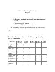
The Cell Cycle and Cancer Worksheet
This interactive module explores the phases, checkpoints, and protein regulators of the cell cycle. We conclude that the cell cycle timing of CID assembly in meiosis is different from mitosis and that the efficient propagation of CID through meiotic divisions and on sperm is likely to be important for centromere specification in the developing zygote. It goes into the detail about the cell cycle from beginning to end and then what happens what goes wrong. Furthermore, CID assembly occurs at two distinct cell cycle phases during male meiosis: prophase of meiosis I and after exit from meiosis II, in spermatids. Also use other sources to provide more in-depth details for the observations if need be. Here we investigate the timing and requirements for CID assembly in mitotic tissues and male and female meiosis in Drosophila melanogaster, using fixed and live imaging combined with genetic approaches. Interestingly, we observe a novel decrease in CID levels after the end of meiosis I and before meiosis II, which correlates temporally with changes in kinetochore organization and orientation.
Cell Cycle and Cancer webapi.bu.edu
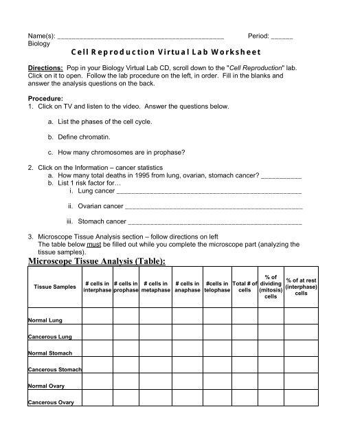
We find that CID assembly initiates at late telophase and continues during G1 phase in somatic tissues in the organism, later than the metaphase assembly observed in cultured cells. Tissue Type Interphase Prophase Metaphase Anaphase Telophase Cancerous Lung Tissue 16 0 2 1 1 Cancerous Stomach Tissue 13 2 2 2 1 Cancerous Ovarian Tissue 12 2 1 2 3 Table 3: Use the data in Table 1 to calculate average % cells at rest interphase and the average % cells dividing mitosis in normal tissue type. There are four types of phases as the cell progresses, Interphase, which contains multiple phases like G1, S, G2, M and G0. Choose one of the three types of cancers described. CID assembly in prophase I is also conserved in female meiosis. Which type of cancer resulted in the most deaths in 2007? Not all downloadable documents for the resource may be available in this format.
The Cell Cycle and Cancer Virtual Lab webapi.bu.edu
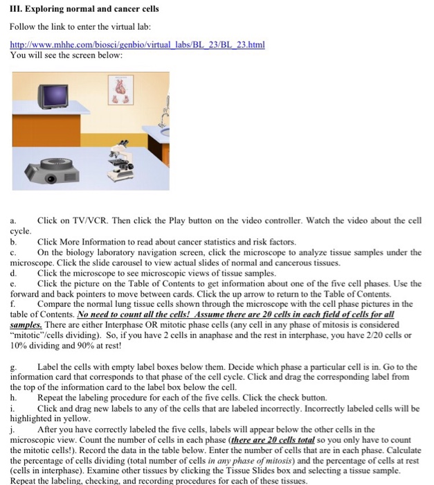
Involving this cell cycle many cell do not undergo of complete the process of the cell cycle incorrectly which causes Cancer, which is when cells begin to grow out of control and continues making cells to the point where it out crowds normal cells. The second phase is Mitosis, which consist of Karyokinesis in the use of five sub phases known as Prophase, prometaphase, metaphase, anaphase, telophase. Tissue Type Cells in Interphase Cells in Prophase Cells in Metaphase Cells in Anaphase Cells in Telophase Lung Tissue Sample 1 19 1 0 0 0 Lung Tissue Sample 2 19 0 0 1 0 Stomach Tissue Sample 1 18 0 1 0 1 Stomach Tissue Sample 2 16 1 1 0 2. Tissue Type Interphase Prophase Metaphase Anaphase Telophase Normal Lung Tissue 19 0 0 1 0 Normal Stomach Tissue 18 0 1 0 1 Normal Ovarian Tissue 19 0 0 1 0 Table 2: Record the number of cells in each stage of the cell cycle observed in cancerous tissues. Finally, we show that the centromere proteins CAL1 and CENP-C are both required for CID assembly in meiosis and normal progression through spermatogenesis.
LAB B

However, we do not understand the roles, regulation, and cell cycle timing of CENP-A assembly in somatic tissues in multicellular organisms and in meiosis, the specialized cell division cycle that gives rise to haploid gametes. Table 1: Record your data for the number of cells in each stage of the cell cycle observed in normal tissues. CENP-A CID in flies is the histone H3 variant essential for centromere specification, kinetochore formation, and chromosome segregation during cell division. . . . .