Tissue level of organization. Glossary: The Tissue Level of Organization 2022-10-26
Tissue level of organization Rating:
9,8/10
1568
reviews
The tissue level of organization refers to the way in which cells are organized into functional units within the body. There are four main types of tissues in the body: epithelial, connective, muscle, and nervous. Each type of tissue performs specific functions and plays a crucial role in maintaining the overall health and function of the body.
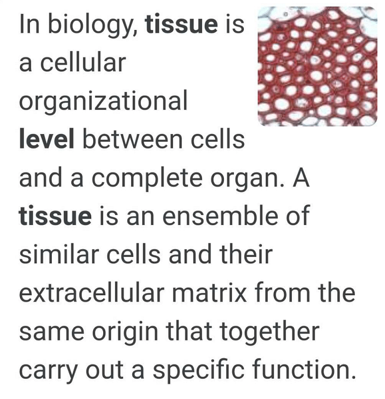
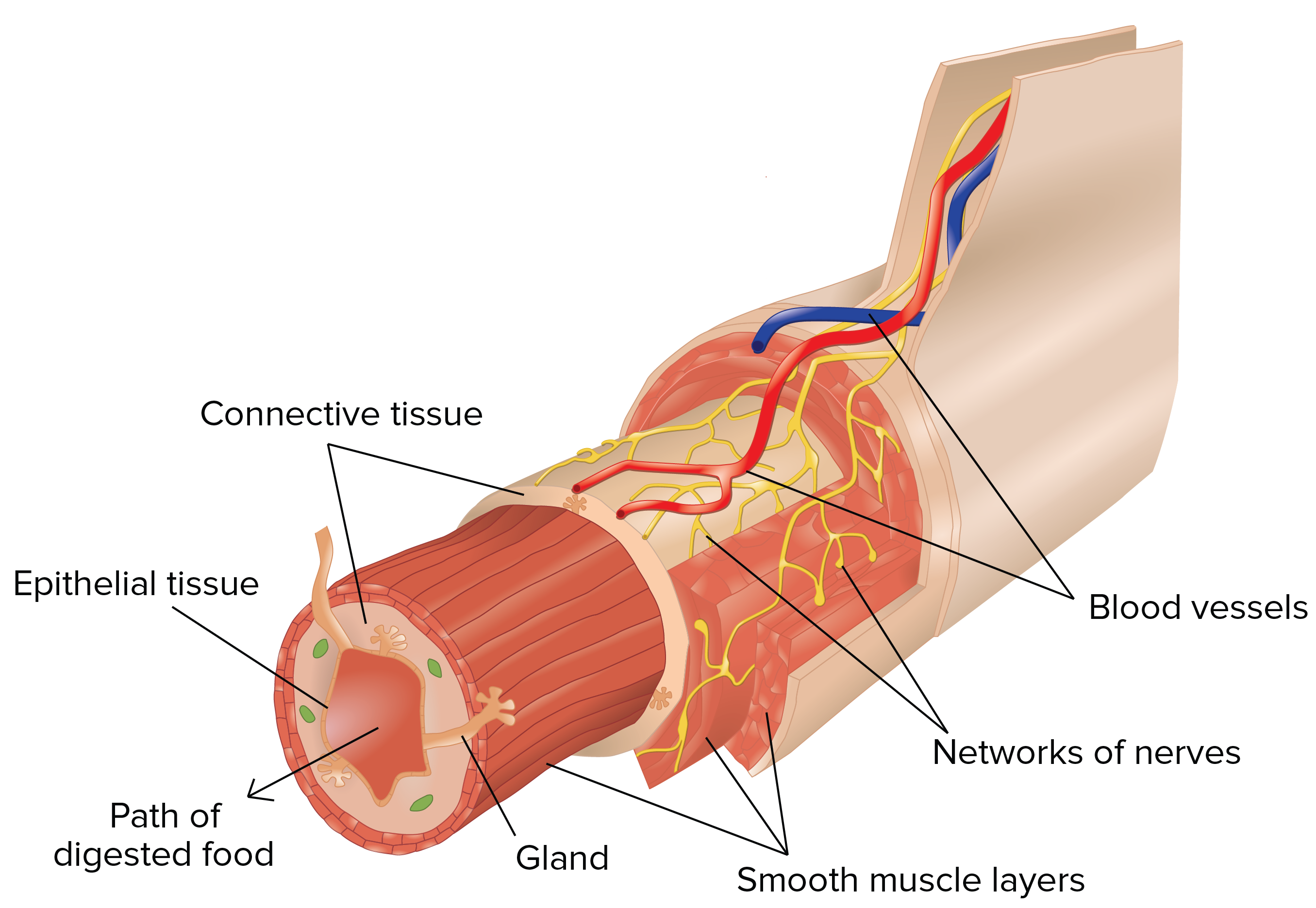
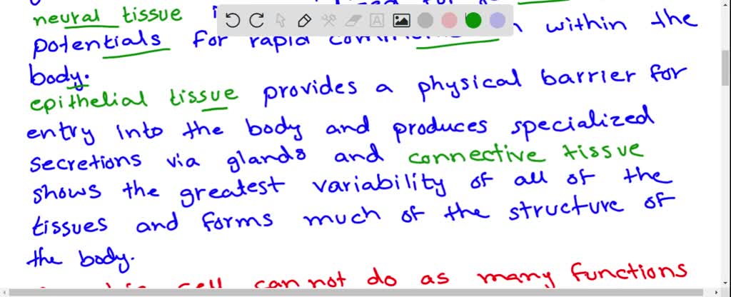
Epithelial tissue forms the outer layers of the body and lines internal organs, blood vessels, and cavities. It functions to protect the body from physical damage, infection, and dehydration. Epithelial tissue is classified based on the number of layers and the shape of the cells. Simple epithelial tissue has a single layer of flat or cuboidal cells, while stratified epithelial tissue has multiple layers of cells that are arranged in a stack-like fashion.
Connective tissue provides support and protection to the body. It is made up of cells that are separated by a matrix, which is a gel-like substance that fills the spaces between the cells. There are several types of connective tissue, including bone, cartilage, and blood. Bone tissue is hard and rigid, and it provides structural support for the body. Cartilage tissue is flexible and resilient, and it is found in joints, the nose, and the ears. Blood tissue is a fluid connective tissue that transports oxygen, nutrients, and hormones throughout the body.
Muscle tissue is responsible for movement and the generation of heat. There are three types of muscle tissue: skeletal, smooth, and cardiac. Skeletal muscle is attached to bones and is responsible for voluntary movement. Smooth muscle is found in the walls of internal organs and blood vessels, and it functions to regulate the size and shape of these structures. Cardiac muscle is found in the heart and is responsible for pumping blood throughout the body.
Nervous tissue is responsible for transmitting and processing information within the body. It is made up of neurons, which are specialized cells that transmit nerve impulses, and glial cells, which support and protect the neurons. The nervous system is divided into the central nervous system, which consists of the brain and spinal cord, and the peripheral nervous system, which consists of the nerves that extend throughout the body.
In summary, the tissue level of organization refers to the way in which cells are organized into functional units within the body. Each type of tissue performs specific functions and plays a crucial role in maintaining the overall health and function of the body.
Tissue Level of Organization
Cer-tain macrophages may be fixed to a site fixedmacrophages , as found in the liver and spleen. Platelets Platelets are the smallest cells present in the blood; they help stop bleeding at the time of injury. Reduced range of motion, ischemic pain, and loss of sensation and voluntary control are some of the negative outcome of adhesions. Connective tissue, such as bone, blood and fat, appear to be different from each other, but they have some common features that place them under this classification. . Be sure you can identify a macrophage and not just a bunch of cells superimposed upon one another. Other cells, such as those in the skin, are bound to-gether by desmosomes.
E areolar connective tissue, adipose connective tissue, and dense connective tissue. A stratified squamous epitheliumhas many lay-ers. Then use your text and atlas to review the diagnostic features of each connective tissue cell present in the micrograph. Macrophages can be seen also in the subcapsular sinus the lighter staining area just under the capsule at the periphery of the lymph node. When the bladder is empty, the cells appear multilayered. The micrograph that opens this chapter shows the high degree of organization among different types of cells in the tissue of the cervix. The epithelium secretes mucous in these re-gions.
Usually clear and colorless, it has the consistency of thick syrup. Microphages Microphages are other types of white blood cells neutrophils and eosinophils that are attracted to sites of injury and inflammation. Fibroblasts Fibroblasts are the most abundant cells. Although collagen fibers mostly fill the view, there are numerous elastic fibers, which provide the elasticity essential for the function of the tissue. Type I reactions, also called anaphylactic reactions, are something you will learn about while studying the immune system later on, but for now, here is a quick explanation. Note the alternating layers of fibroblasts and collagenous fiber bundles. The squamous, cuboidal, and columnar ep-ithelium may be simple or stratified.
Sim-ple squamous epithelium see Figure 1. Proteins, nutrients, waste products, hormones, and electrolytes are dis-solved in the plasma. Unlike epithelia, the cells in connective tissue are scattered. These cells contain essentially the same internal structures yet they vary enormously in shape and function. In the nucleus, areas of euchromatin and heterochromatin can easily be identified. Together, all these differentiated cells are able to fulfill the needs of the body. Instead, some cells become specialized to do specific functions.
Chapter 4 The Tissue Level of Organization Flashcards
Cells may be bound to each other by fusion of cell membranes to form tight junctions. These connections are strong and help to maintain the cell layers in sheets. Make sure you can see the difference between cross sectioned and longitudinally sectioned collagenous fibrils. As it is largely made up of nonliving material, its fluid crystal state can be manipulated to a large ex-tent by application of heat, cold, stretch, and activity. Structure As the major function of epithelia is to form a barrier, they are found in layers, with individual cells bound to adjacent cells, unlike other tissues that may be found scattered individually in the extracellular ma-terial. The simplest level is the chemical level, which includes tiny building blocks such as atoms. Organs: Made of Tissues An organ is a structure that is composed of at least two or more tissue types and performs a specific set of functions for the body.
Classification Connective tissue may be classified as connectivetissue proper, fluid connective tissue,and sup-porting connective tissue. You can see one large lipid droplet in the cytoplasm of each cell. These cells are important in removing all kinds of debris from the body as well as playing a major role in the immune response. The fibers may be colla-gen fibers, reticular fibers,or elastic fibers. In bone, in addition to the fibers, insoluble calcium salts are deposited in the ground substance. As they have a phagocytic function, removing pathogens and cell debris, macrophages usually contain abundant primary and secondary lysosomes. Muscle tissue is classified into three types according to structure and function: skeletal, cardiac, and smooth.
Neutrophils generally enter tissues in large numbers only in response to a disease stimulus. It is also found in the epiphyseal plate the region where bone growth occurs. The simple epithelium lining blood vessels and heart are called endothelium. Simple Epithelium Simple epithelium has only one layer of cells over the basement membrane. These cells contain essentially the same internal structures yet they vary enormously in shape and function. The property of cartilage depends on the type and proportion of protein fibers scattered in the matrix.
Fluid Connective Tissue Blood and lymph are examples of fluid connective tis-sue. They may be arranged randomly, forming sheets e. Strength is provided by the presence of numerous fibers in the ground substance. By having a watery ground sub-stance, fluid connective tissue, such as blood, is formed. Almost all epithelia have a good nerve supply, which enables them to sense changes in the environ-ment and convey that information to the brain for suitable action.
Ten-dons and ligaments, which withstand a lot of force as muscle contracts, are made up almost entirely of colla-gen. Please only use it after you've completed the problem set yourself. The layers do not contain any blood vessels and one surface of the cells lines the cavity of the organ. If all other tissue was removed, connective tissue would form the three-dimensional framework of the body, much like cellulose in plants. Dense connective tissue has a shiny, white appearance. You will study neutrophils in much greater detail in other sequences and in your histopathology course, but it is useful for now to at least be able to recognize them in various tissues and organs.