The neuromuscular junction review sheet 14. The Neuromuscular Junction Review Flashcards 2022-10-31

The neuromuscular junction review sheet 14 Rating: 5,6/10 1382 reviews

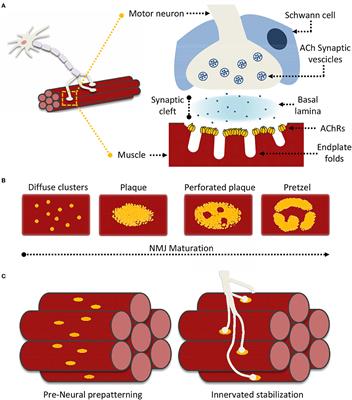
The neuromuscular junction is the point of communication between a motor neuron and a skeletal muscle fiber. It is a chemical synapse, meaning that it uses chemical signaling to transmit the signal from the neuron to the muscle.

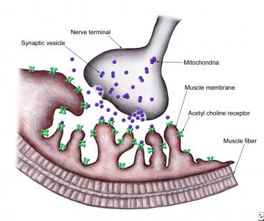
The process of signaling at the neuromuscular junction begins when an action potential, or nerve impulse, reaches the axon terminal of the motor neuron. This causes the release of the neurotransmitter acetylcholine (ACh) from the terminal into the synapse, or gap, between the neuron and the muscle.

ACh then diffuses across the synapse and binds to receptors on the muscle fiber, called nicotinic receptors. This binding causes the receptors to open ion channels, allowing ions such as sodium and potassium to flow into the muscle fiber. This influx of ions causes a change in the muscle fiber's membrane potential, called a depolarization.

If the depolarization reaches a certain threshold, it will trigger an action potential in the muscle fiber, which will then cause the muscle to contract. This process of signaling at the neuromuscular junction is known as neuromuscular transmission.

There are several factors that can affect the efficiency of neuromuscular transmission. One of these is the concentration of ACh at the axon terminal. If there is too little ACh, the signal may not be strong enough to trigger an action potential in the muscle fiber. On the other hand, if there is too much ACh, it can lead to excessive muscle activity, such as cramping or spasms.

Another factor that can affect neuromuscular transmission is the presence of drugs or toxins that interfere with the signaling process. For example, curare is a toxin that blocks the nicotinic receptors on the muscle fiber, preventing ACh from binding and inhibiting muscle contraction.

In summary, the neuromuscular junction is an important point of communication between the nervous system and the skeletal muscles. It relies on the release and binding of the neurotransmitter ACh to transmit the signal from the neuron to the muscle and trigger muscle contraction. Various factors, such as the concentration of ACh and the presence of drugs or toxins, can affect the efficiency of this process.

Mechanisms Regulating Neuromuscular Junction Development and Function and Causes of Muscle Wasting

the neuromuscular junction review sheet 14

In addition, old muscle regenerates poorly upon injury due to decreased regenerative capacity of aged satellite cells see review by Ref. Myoblasts fuse to form primary myotubes only after they reach the full extent of the diaphragm and are subsequently innervated by multiple motor axons Ab. Embryonic to perinatal stages Motor nerves reach the diaphragm at E12. J Physiol 339: 355—377, 1983. Hum Mutat 33: 1474—1484, 2012. Mapping sites responsible for interactions of agrin with neurons.

Next

Neuromuscular Junction

the neuromuscular junction review sheet 14

C: in mature muscle, each muscle fiber is innervated by one motor neuron. Effects of purified recombinant neural and muscle agrin on skeletal muscle fibers in vivo. Only higher-volume release from large numbers of vesicles can produce the voltage changes that forward a nerve impulse. While it has been proposed that expression of autophagy genes Tsc1 cause accelerated muscle atrophy upon denervation rather than sparing C, see also A. Immature Schwann cells differentiate into myelinating and nonmyelinating Schwann cells, depending on whether they associate with large or small axons or with nerve terminals terminal Schwann cells , respectively. Muscle Nerve 17: 820—821, 1994. Interestingly, consistent with the complementary roles of synaptic fold depth and density, and of synaptic growth for maintaining a high safety factor of neuromuscular transmission, these two parameters are inversely correlated in different species 4.

Next

Neuromuscular junction: Parts, structure and steps

the neuromuscular junction review sheet 14

Utrophin links the entire complex to the F-actin cytoskeleton. J Neurosci 25: 9949—9959, 2005. One of the challenges with this parameter is the fact that muscle mass can be measured with different methods. Like humans, aged mice and rats older than 24 mo show loss of skeletal muscle mass. B: high magnification of the neuromuscular junction. The enrichment of myogenic cells in the center of the diaphragm is reflected by a central, dense band of nuclei that express high levels of myogenin Ba.

Next

The Neuromuscular Junction Review Flashcards

the neuromuscular junction review sheet 14

At this stage, the primary and the secondary myotubes are electrically coupled Ba. Fasting-induced muscle atrophy is based on the requirement to generate free amino acids for protein synthesis in the absence of amino acids supplied by the food. Genetic forms of neuromuscular junction disorders span diseases such as muscular dystrophy and the group of congenital myasthenic syndromes. One size may not fit all: anti-aging therapies and sarcopenia. Sarcopenia Is a Multifactorial Syndrome of Old Age The term sarcopenia 2. Furthermore, it should be noted that we will not discuss all the pathways that have been implicated in neuromuscular disorders by genetic experiments, as the function of many of the respective gene products remains obscure.

Next

the neuromuscular junction review sheet 14

Vesicles transport acetylcholine into the synaptic cleft. Thus they should be adopted with caution to the human situation; for example, the clinical manifestation of a defect may be different for rodents and humans with their different loads on muscles associated with the different postures. Polyneuronal innervation of skeletal muscle in new-born rats and its elimination during maturation. Muscle Atrophy and Muscle Wasting Can Be Triggered by Many Factors Muscle atrophy can also be triggered by mechanical unloading e. Rapid and reversible effects of activity on acetylcholine receptor density at the neuromuscular junction in vivo. As life expectancy is increasing, loss of muscle mass during aging, called sarcopenia, has emerged as a field of high medical need.

Next

the neuromuscular junction review sheet 14

Regulation of Chrn gene expression at the synapse Several molecules from the nerve have been implicated in mediating the neural regulation of synapse-specific Chrn and Musk gene expression in fundamental nuclei. The reader should keep in mind, however, that most of the mechanistic data available on this topic stem from experiments with rodents. ErbB proteins are receptor tyrosine kinases comprising ErbB2, ErbB3, and ErbB4 isoforms, which are encoded by distinct genes. Constructing and decoding unconventional ubiquitin chains. A muscle might fasciculate spasm for some time. For example, in the adult rat diaphragm, they are the source of up to 80% of the muscle fibers Because of this problem, important concepts have also been derived from the study of so-called ectopic endplates formed in the adult animal.

Next

the neuromuscular junction review sheet 14

Protein ubiquitination involves the sequential activation and conjugation of ubiquitin monomers by E1 and E2 enzymes. Absence of acetylcholinesterase at the neuromuscular junctions of perlecan-null mice. Voltage-gated sodium channels and ankyrinG occupy a different postsynaptic domain from acetylcholine receptors from an early stage of neuromuscular junction maturation in rats. This means that the motor end plate will also be located near the midpoint of the muscle fiber. The functionality of such complexes relies on the ability of the cullins to link the polyubiquitin-loaded E2 with the specific substrate to be degraded B. Development of phrenic motoneuron morphology in the fetal rat.

Next

the neuromuscular junction review sheet 14

In vivo and in vitro analysis of electrical activity-dependent expression of muscle acetylcholine receptor genes using adenovirus. Acetyl choline is released into the synaptic cleft. In these latter conditions, innervation of the muscle remains intact, but muscle activity is decreased. The majority of muscle fibers in the adult muscle are derived from secondary myotubes. J Cell Sci 123: 3944—3955, 2010. B: at late embryonic development from E14. Rapsyn may function as a link between the acetylcholine receptor and the agrin-binding dystrophin-associated glycoprotein complex.

Next