Right common iliac artery. Iliac Artery 2022-10-15
Right common iliac artery Rating:
8,8/10
213
reviews
The right common iliac artery is a major blood vessel in the human body that carries oxygenated blood from the aorta to the lower half of the body. It is one of two common iliac arteries, the other being the left common iliac artery, which carries blood to the left side of the body.
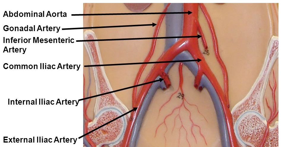
The right common iliac artery begins at the level of the fourth lumbar vertebra, where it branches off from the aorta. From there, it travels downward and to the right, passing through the pelvis and into the right leg. Along its course, the right common iliac artery gives off several smaller branches, including the internal iliac artery and the external iliac artery.
The internal iliac artery supplies blood to the muscles and organs of the pelvis, including the bladder, uterus, and rectum. The external iliac artery, on the other hand, supplies blood to the muscles and skin of the lower extremities, including the thighs, knees, and ankles.
Problems with the right common iliac artery can have serious consequences for a person's health. One such problem is an arterial blockage, also known as arterial occlusion, which can lead to reduced blood flow to the lower half of the body. This can cause symptoms such as leg pain, numbness, and weakness, and if left untreated, it can lead to more serious complications such as tissue death and amputation.
Treatment for problems with the right common iliac artery typically involves the use of medications to widen the artery and improve blood flow, as well as lifestyle changes such as quitting smoking and adopting a healthy diet. In more severe cases, surgical procedures may be necessary to repair or bypass the damaged artery.
In conclusion, the right common iliac artery is a vital blood vessel that plays a crucial role in supplying oxygenated blood to the lower half of the body. Problems with this artery can have serious consequences for a person's health, but with proper treatment, it is possible to manage and prevent these problems from occurring.
Common iliac vein: Anatomy and drainage
Bodytomy provides a labeled iliac artery diagram to help you understand the anatomy and function of the common iliac. Â Anatomy of the Human Body. The union of the internal and external iliac veins creates the common iliac vein, while the inferior epigastric vein drains into the external iliac vein and anastomoses from the superior epigastric vein. These smaller channels of the iliac arteries divide into even smaller arteries to reach more of your lower body. These vessels run parallel with their venous counterparts, the internal and external iliac veins, which join to form the inferior vena cava. Rupture of iliac artery aneurysms is reported to carry a 50% to 70% mortality rate and because the incidence of rupture is as high as 50%, it is imperative that diagnosis be early and intervention prompt 11. The largest artery in the body the aorta divides into the common iliac arteries.
Origin of Common Iliac and Internal and External Iliac Arteries Internal iliac artery branches The internal iliac artery divides into anterior and posterior trunks at the level of the superior border of the greater sciatic foramen. It supplies the musculature of the thigh. The deep iliac circumflex artery arises from the side of the external iliac. The superior vesical artery is also responsible for the blood supply to the adjacent ureter and vas deferens. Arterial occlusion is the blockage of the artery by a clot or plaque, which can lead to reduced blood flow to the lower extremities and potentially cause serious complications such as amputation or gangrene. This vessel makes a substantial contribution to the blood supply of muscles and skin in the gluteal region and also supplies branches to adj acent muscles and bones of the pelvic walls.
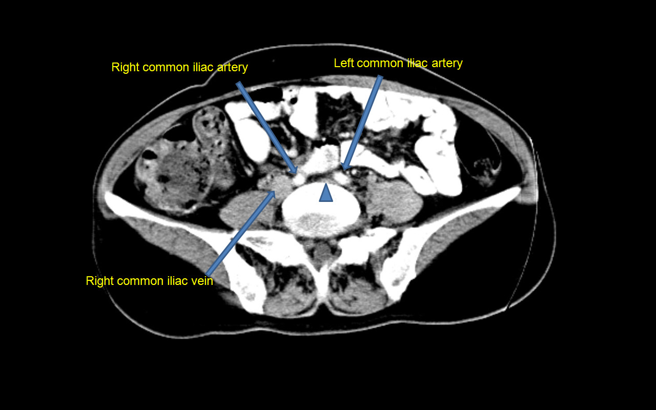
A rapidly growing aneurysm about to rupture burst can be tender and very painful when pressed. The vaginal artery in women is the equivalent of the inferior vesical artery in men and, descending to the vagina, supplies branches to the vagina and to adjacent parts of the bladder and rectum. The 3 major branches of the posterior division include the iliolumbar artery, lateral sacral artery, and the superior gluteal artery, the large terminal branch of the posterior division, which passes posteriorly and runs between the lumbosacral nerve trunk and the anterior ramus of the S1 nerve. These arteries are known as common iliac arteries and there are two such arteries, one on each side of the lower abdomen. Inferior vena cava - ventral view Inferior vena cava filter- If someone had recurrent deep vein thrombosis and is at risk of recurrent pulmonary embolism or is non compliant with anticoagulant medication a small filter can be passed through the femoral vein, through the common iliac vein and into the infrarenal inferior vena cava through an image guided procedure.
Iliac artery aneurysm: Types, causes, symptoms, diagnosis, and treatment
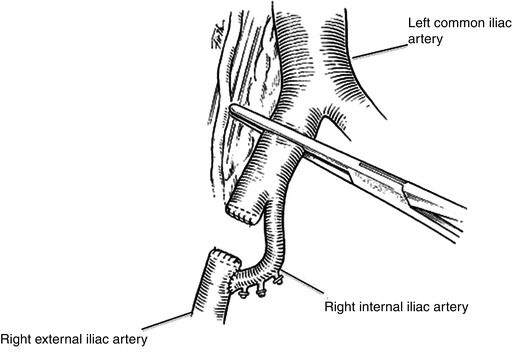
The femoral artery supplies blood to the thigh, the popliteal artery supplies the knee area, and the anterior and posterior tibial arteries supply the area below the knee, including the feet and toes. Instead, a graft is inserted into the aorta to strengthen it. They give off small branches to surrounding structures before dividing into the This article will discuss the Key facts about the common iliac artery Origin Abdominal aorta Branches Internal and external iliac arteries, branches to peritoneum, psoas major muscle, ureter, and adjacent nerves Supply Lower extremity via external iliac artery , pelvis and its viscera via internal iliac artery , peritoneum, psoas major muscle, ureter, surrounding tissues, lymph nodes and nerves The common iliac artery arises from the aortic bifurcation at the L4 level. An antegrade approach to common iliac artery recanalization may also be considered, from either the contralateral groin or an upper limb approach. Although the chances of experiencing iliac artery aneurysm are low, if affected, the symptoms and complications can become quite severe. The aorta goes through the chest and abdomen.
Common Iliac Artery: Anatomy, Function, and Significance
The superior gluteal artery passes between the lumbosacral trunk and the ventral ramus of S1. Common iliac vessels inside a cadaver: The common iliac vein has a larger diameter compared to the artery of the same name. On the other hand, if osteotomies are necessary, they are more risky on a vascular level than the fibula. The common iliac veins formed by the union of external and internal iliac veins lie medial to the corresponding arteries at a deeper plane. Signs and symptoms also depend on whether the aneurysm has ruptured burst or is affecting other parts of the body. This can lead to a stroke. A cardiothoracic surgeon does surgery on the heart, lungs, and other organs and structures in the chest, including the aorta.
Anatomy and Function of the Common Iliac Artery With Labeled Diagrams
There appears to be no difference in blood flow between the two uterine arteries in relation to which ovary contains the dominant follicle. This is an important variation to be aware of in diagnostic procedures involving the vessel e. The right common iliac artery divides into the right internal and external iliac arteries. . The internal iliac artery supplies blood to the pelvis and reproductive organs, while the external iliac artery supplies blood to the lower extremities. Follow a healthy diet, get regular exercise, and keep your cholesterol at a healthy level to also help prevent aneurysms or their complications. It is at high risk for damage during both arthroscopic and open anterior approaches to the hip.
In conclusion, the common iliac artery is a vital blood vessel in the human body that supplies oxygen and nutrients to the lower extremities, pelvis, and buttocks. It supplies blood to the greater sciatic foramen, which is located above the upper border of piriformis. Among the structures it supplies are the erectile tissues of the clitoris and the penis. The risk for abdominal aortic aneurysms increases as you get older. Percutaneous embolization of an internal iliac artery aneurysm: technical considerations and literature review. The aorta passes down through the chest, where it is called the thoracic aorta, and continues into the abdomen, where it is called the abdominal aorta. The most important parts are the ovaries, fallopian tubes, uterus and vagina.
They emerge superior to the piriformis and then terminate in the gluteus medius and gluteus minimus muscles. A small branch of the obturator artery provides blood to the periosteum of the back of the pubis. Overlying these neurovascular structures is a reflected portion of the parietal peritoneum and the obturator internus muscle. The left and right common iliac arteries are the terminal branches of the abdominal aorta. Common iliac artery As the abdominal aorta progresses downwards into the abdomen, it divides into two large arteries. The extra pelvic veins include the superior and inferior gluteal veins, the obturator drains the lateral The common iliac veins unite with the common iliac vein of the contralateral side slightly at the right side of vertebral level L5.
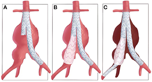
Guidance for Surveillance of Isolated Common Iliac Artery and Small Abdominal Aortic Aneurysms
Palpation of the sciatic notch and the posterior column can help to prevent the placement of proud screws and further decrease the risk of injury. The two main types of surgery to repair aortic aneurysms are open abdominal or open chest repair and endovascular repair. There were 1,581 patients mean age, 73 years; 78% male and 93% white and nearly 6,000 ultrasounds, with a mean follow-up of 28 months. Iliac artery aneurysm presenting with lower extremity deep vein thrombosis. The common iliac artery is a major blood vessel in the human body that supplies oxygen-rich blood to the lower extremities, pelvis, and buttocks. Atherosclerotic disease cholesterol buildup in arteries may also lead to the formation of some aneurysms 21.
Iliac Artery: What Is It, Location, Anatomy and Function
During his active practice he served as the head of the Dept. Sara Paterson-Brown, in Basic Science in Obstetrics and Gynaecology Fourth Edition , 2010 The common iliac arteries The common iliac arteries diverge from in front of the fourth lumbar vertebra and then divide into internal and external iliac arteries in front of the sacroiliac joint. The information we provide is grounded on academic literature and peer-reviewed research. The profundus femoris artery, which is also known as the deep femoral artery, is the first branch of the common femoral artery. Certain factors put you at higher risk for an aortic aneurysm. You can make diet and lifestyle changes to keep the iliac arteries open and healthy. It provides blood to the uterine tube and joins the tubal branch of the ovarian artery.