Pancreas anatomy. Pancreas: Anatomy and Functions 2022-11-06

Pancreas anatomy Rating: 9,9/10 1085 reviews

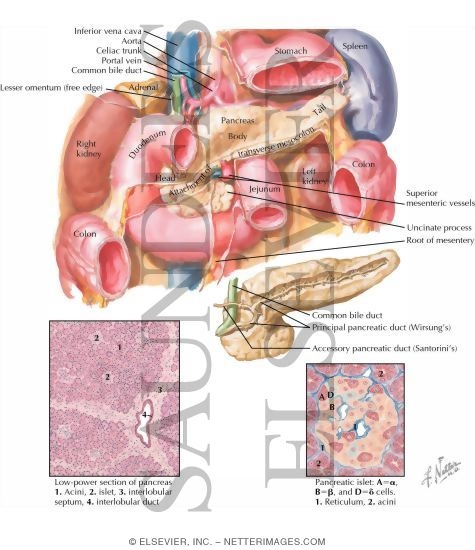
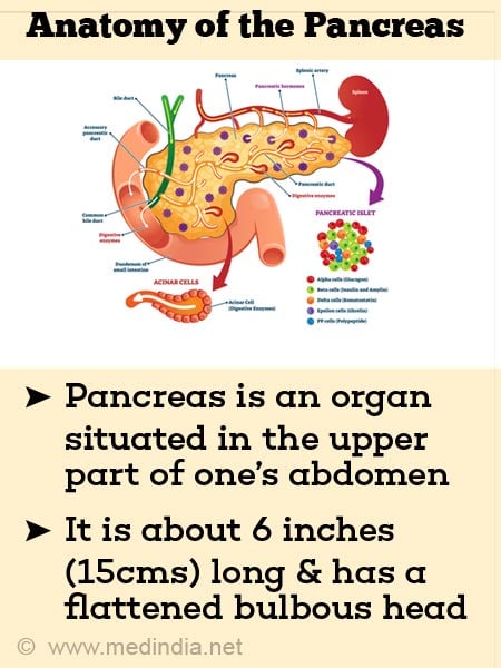
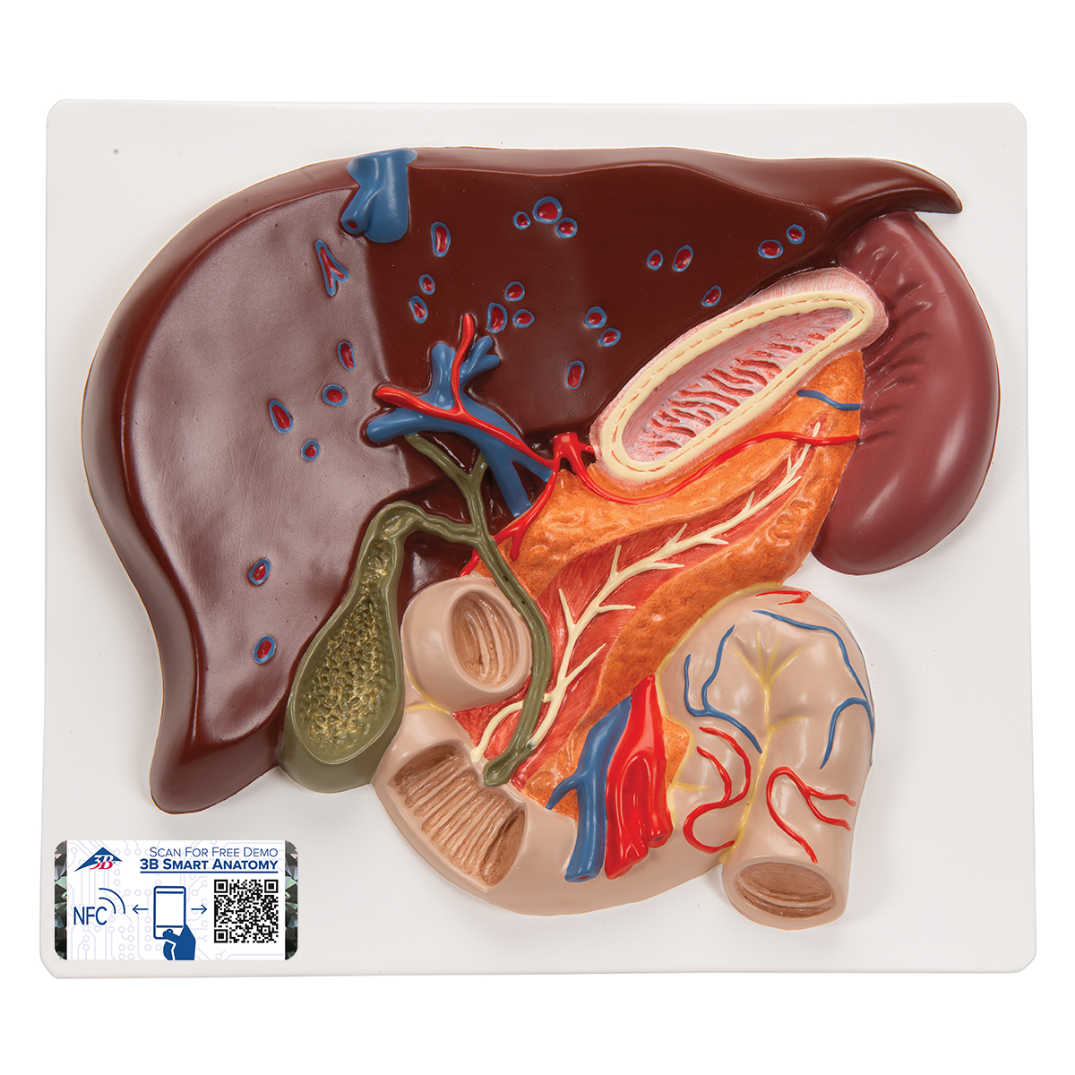
The pancreas is a gland located in the abdomen, behind the stomach. It is about 6 inches long and shaped like a flat pear. The pancreas has two main functions: it produces hormones such as insulin and glucagon, which help regulate blood sugar levels, and it produces enzymes that aid in digestion.

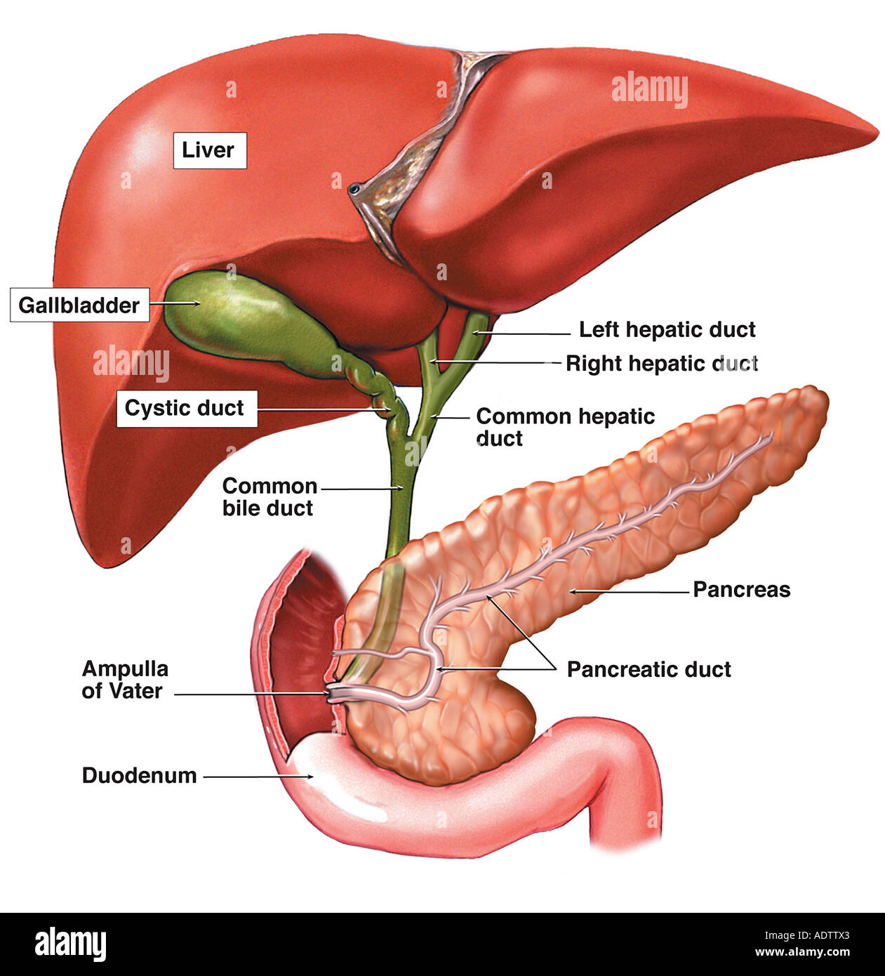
The pancreas has two main parts: the exocrine portion, which makes up about 80% of the gland, and the endocrine portion, which makes up the remaining 20%. The exocrine portion of the pancreas produces and releases digestive enzymes into the small intestine through a system of ducts. These enzymes help to break down fats, proteins, and carbohydrates in the food we eat.

The endocrine portion of the pancreas, also known as the islets of Langerhans, produces hormones that regulate blood sugar levels. The main hormone produced by the pancreas is insulin, which helps to lower blood sugar levels by helping cells in the body use glucose for energy. Another hormone produced by the pancreas is glucagon, which helps to raise blood sugar levels by stimulating the liver to convert stored glycogen into glucose.

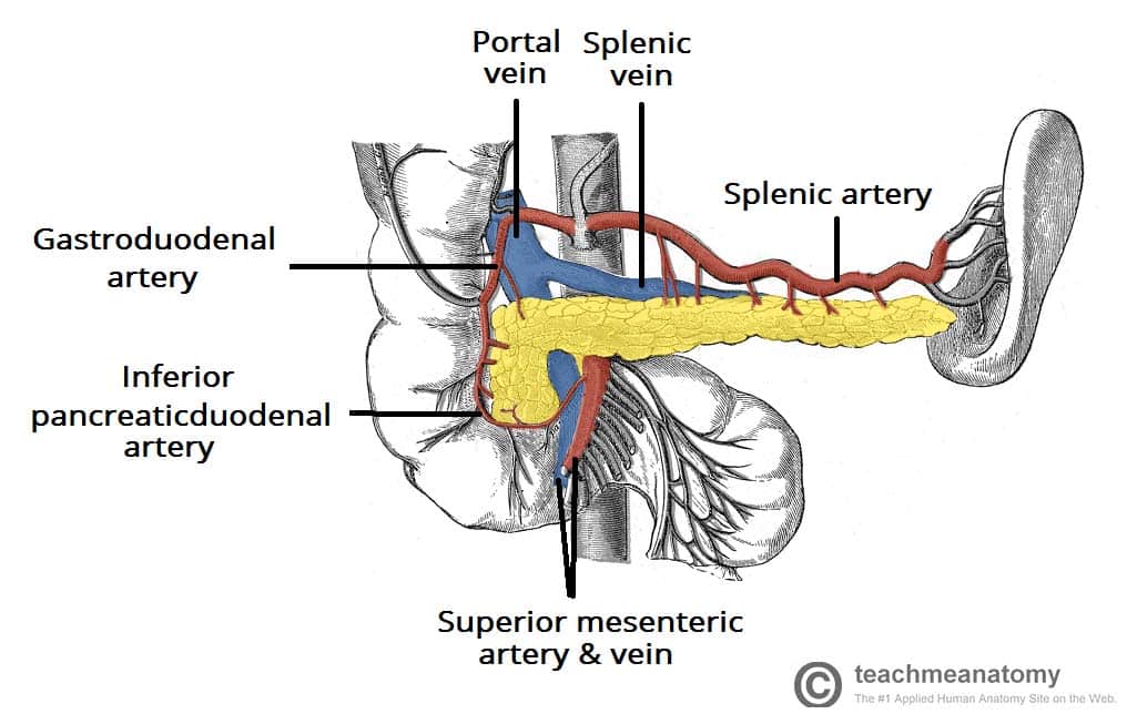
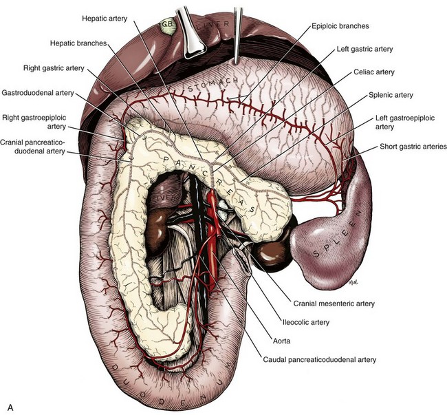
The pancreas also has a rich blood supply, with arteries and veins that bring oxygen and nutrients to the gland and carry away waste products. The main artery supplying blood to the pancreas is the splenic artery, which branches off from the celiac artery. The veins of the pancreas drain into the portal vein, which carries blood from the digestive organs to the liver.

Problems with the pancreas can lead to serious health issues. One common problem is pancreatitis, which is inflammation of the pancreas. This can be caused by a blockage of the ducts that carry digestive enzymes, or it can be caused by excessive alcohol consumption. Another problem with the pancreas is diabetes, which is a disease in which the body does not produce enough insulin or does not use it effectively. This can lead to high blood sugar levels and a variety of related health problems.

In conclusion, the pancreas is a vital gland located in the abdomen that has both exocrine and endocrine functions. It produces hormones that regulate blood sugar levels and enzymes that aid in digestion. Problems with the pancreas can lead to serious health issues such as pancreatitis and diabetes.

Pancreas transplant

pancreas anatomy

Type 1 diabetes is an autoimmune disease affecting the beta cells of the pancreas. If you do, you'll need to return to the hospital for treatment with intensive anti-rejection medications. Endocrine pancreas, the portions of the pancreas the islets that make and secrete insulin, glucagon, somatostatin and pancreatic polypeptide into the blood. Role of actin in regulated exocytosis and compensatory membrane retrieval: insights from an old acquaintance. Acinar cells stain blue at their base because of the high content of RNA and the presence of nuclei. This location is one of the most protected sites of the body, making it resistant to injury, but hard to examine. Islets vary greatly in size; ~70% are in the size range of 50-250 μm in diameter in humans with an average in the range of 100-150 μm 4.

Next

Pancreas Anatomy and Function

pancreas anatomy

Often, people with type 2 diabetes are overweight or obese, although weight is not the only risk factor. Accessed April 21, 2019. It is secreted by alpha cells, which make up 20% of cells in the pancreas. The head lies near the duodenum and the tail extends to the hilum of the spleen.   As a result, patients feel constant pain in the upper abdomen that radiates to other parts of the body.

Next

El páncreas: Anatomía y funciones

pancreas anatomy

Use of the terms left and right can be problematic. This includes acinar and duct cells with associated connective tissue, vessels, and nerves. If you're waiting for a donated pancreas, make sure the transplant team knows how to reach you at all times. Lymph nodes draining the pancreas. Colloquium Series on Integrated Systems Physiology: From Molecule to Function. It is a medication made of killed or weakened cells, organisms or manufactured materials, which is used to boost the body's immune system.

Next

Normal Pancreas

pancreas anatomy

It results from the failure of fusion of dorsal and ventral pancreatic 1,3. The connective tissue component of the duct wall becomes progressively thinner as the ducts branch and become narrower. The pancreas is about the size of the half of your hand that includes the index and third fingers excluding the thumb. Image by Jennifer Parsons Brumbaugh; used with permission of the publisher 6. Biochem Biophys Res Commun.


Next

Pancreatic ducts

pancreas anatomy

Thinner 1 μm sections of plastic embedded tissues prepared for electron microscopy may also be used for light microscopy and a few such sections are also illustrated. The dorsal pancreatic anlage gives rise to a portion of the head, the body, and tail of the pancreas including a major duct that is continuous through the three regions. Mouse and human islets stained for glucagon Immunoperoxidase. The waiting time depends on your blood group and how long it takes for a suitable donor — one whose blood and tissue types match yours — to become available. The endocrine gland consists of the islets of Langerhans. The islands are called the islets of Langerhans.


Next

17.9 The Pancreas

pancreas anatomy

Pancreatic ducts Traveling within the entire pancreatic parenchyma from the tail to the head is the main pancreatic Wirsung duct. It has the potential of invading into the adjacent tissues, spreading to other organs and may eventually lead to the patient's death. These enzymes travel down the pancreatic duct into the bile duct in an inactive form. Embryology and Development The pancreatobiliary anlagen appear at gestation week 5 in the human; fusion of the dorsal and ventral anlagen occurs during week 7 10. The lumen of the ductule contains a pink granular proteinaceous precipitate from pancreatic juice.

Next

Pancreas Anatomy & Diagram

pancreas anatomy

Perforation Disorders in pancreatic structure can lead to holes in the organ, in which case digestive enzymes leak into the abdominal cavity. The upper panel A is visualized from the front, and the lower panel B is seen from the back. Superior and inferior are used in the same context so that they mean toward the head and toward the feet, respectively. This narrow organ lies along the first segment of your small intestine, called the duodenum. These enzymes are secreted into a network of ducts that join the main pancreatic duct. Anatomic variations in the pancreatic and common bile duct systems.

Next

Anatomy and Histology of the Pancreas

pancreas anatomy

M, Sempoux C, Henquin J. Treasure Island FL : StatPearls Publishing. Pancreatic islet transplantation in humans: Recent progress and future directions. Blue zymogen granules are conspicuous in the acinar cells. The narrow end of the pancreas, called the tail, extends to the left side of the body. It also prevents reflux of duodenal contents into the hepatopancreatic duct.

Next

Pancreas (Human Anatomy): Picture, Function, Conditions, Tests, Treatments

pancreas anatomy

Micrograph contributed by James Jamieson. As of 2012 the American Diabetes Association reports that diabetes mellitus has been diagnosed in more than 21 million people in the United States; more than 200,000 of those are children. In type 2 diabetes, cells become resistant to the effects of insulin. Removable tissues include some lymph nodes and parts of the duodenum and stomach that are routinely removed in some surgical treatments for pancreatic cancer. The pancreas secretes enzymes needed for the digestion of food and it produces hormones such as insulin and glucagon which help control blood sugar.

Next

Pancreas: Anatomy, Function, and Treatment

pancreas anatomy

It is worthwhile to mention a few definitions for key terms as used in the context of the pancreas: Exocrine pancreas, the portion of the pancreas that makes and secretes digestive enzymes into the duodenum. Pancreas and islet transplantation in diabetes mellitus. In turn, the body and tail of the pancreas are supplied by pancreatic arteries that stem from the splenic, gastroduodenal, and superior mesenteric arteries. Figures 1A and 1B are two normal human pancreases from autopsies of adults. It's not unusual for pancreas transplant recipients to experience an acute rejection episode within the first few months after the procedure. Pancreas Location and Anatomy The pancreas is elongated in shape and extends horizontally across the upper abdomen.


Next