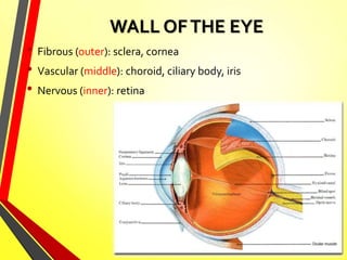
Middle coat of the eyeball. Eyeball: Structure and function 2022-10-16
Middle coat of the eyeball Rating:
5,2/10
946
reviews
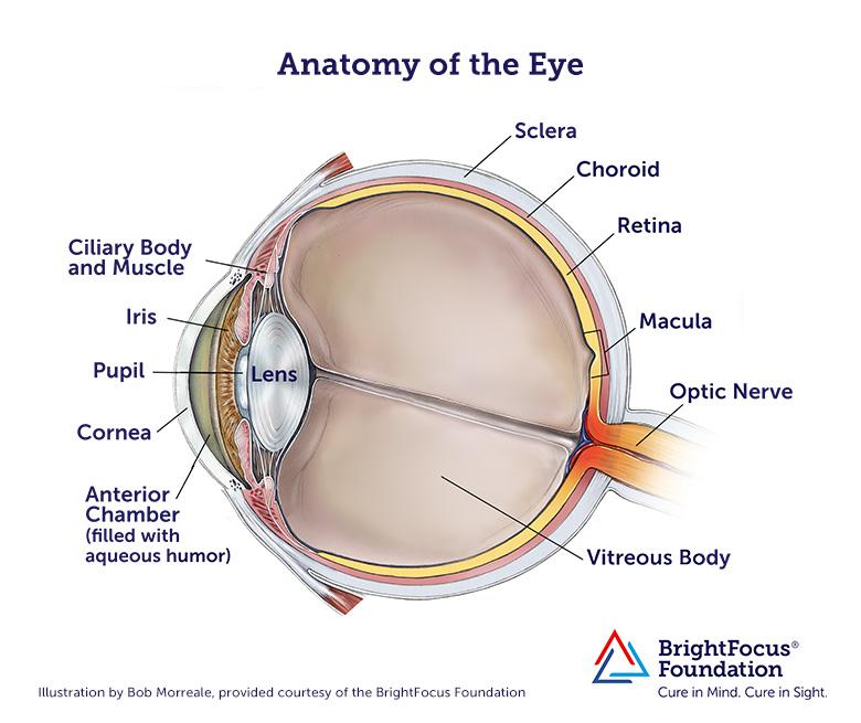
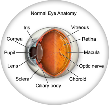
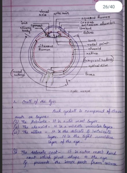
The middle coat of the eyeball, also known as the vascular layer or vascular tunic, is an important layer that provides nourishment to the other layers of the eye and helps maintain its overall health. It is located between the outer fibrous layer and the inner neural layer and is made up of several different structures, including the choroid, ciliary body, and iris.
The choroid is a layer of blood vessels and connective tissue that sits between the sclera (the white outer layer of the eye) and the retina (the inner layer that converts light into neural signals). It is responsible for providing oxygen and nutrients to the retina and for regulating the temperature of the eye. The choroid also contains pigment cells that help absorb excess light and prevent glare.
The ciliary body is a muscular structure located behind the iris (the colored part of the eye). It is responsible for controlling the shape of the lens, which helps to focus light on the retina. The ciliary body also produces aqueous humor, a clear fluid that fills the space between the cornea and the lens. Aqueous humor helps to maintain the shape of the eye and provides nutrients to the cornea and lens.
The iris is the colored part of the eye that surrounds the pupil (the black circle in the center of the eye). It is made up of muscle fibers that control the size of the pupil and allow the eye to adjust to different light conditions. The iris also helps to protect the eye from bright light and debris by constricting the pupil.
Overall, the middle coat of the eyeball plays a vital role in maintaining the health and function of the eye. It provides nourishment and support to the other layers of the eye, helps regulate the temperature and shape of the eye, and helps protect the eye from bright light and debris.
What is the blackish middle coat of the eyeball called?
It is structurally similar to the choroid but lacks the choriocapillaris and the suprachoroidea is less developed. Eyeball Bulbus oculi The eye is a highly specialized sensory organ located within the The average human eye can see around 100 different shades of color and has a resolution that equals 576 gigapixels. Considering the dome shaped surface of the cornea, this structure refracts bends light as it passes through the eye. The pressure with these vessels is higher than the IOP and pressure in most other parts of the body. In the average person, the optic disc carries about 1. A vertical line passing through the fovea centralis divides the retina into the nasal and temporal halves.
Eye Anatomy: 16 Parts of the Eye & Their Functions
It is the thickest corneal layer, comprising around 90% of the thickness of the cornea. The choroid extends from the site of exit of the optic nerve posteriorly, to the ciliary body anteriorly. This layer is absent in the crypts where the aqueous comes into direct contact with the stromal blood vessels. These layers enable the eye to keep its shape. At the same time, the pupil of the other eye also contracts.
The middle coat of the eyeball that contains pigment which ...
The humor flows through the pupil into the anterior chamber of the eye, where it is absorbed into the scleral venous sinus the canal of Schlemm. Â These three layers comprise the circular outline of the eyeball. The macula lutea features a shallow depression in its center, called the fovea centralis. The iris also performs what is known as the " accommodation reflex. The function of the vitreous body is to contribute to the refraction of light, although its dioptric index is significantly smaller than that of cornea and lens. This action requires adjusting the pupil's aperture opening , the shape of the lens, and convergence the ability of the eyes to work together.
The middle coat of the eyeball Codycross [ Answers ]
Fibrous layer Sclera The sclera is an opaque, white, outer layer that surrounds the posterior five-sixths of the eyeball. A horizontal line passing through the fovea further divides the halves into the four quadrants; the upper temporal, lower temporal, upper nasal, and lower nasal. This enables you to see and interpret what you see. Vitreous body Synonyms: Vitreous humour, vitreous humor The vitreous body is the largest structure of the eyeball, occupying the four-fifths of the entire eye. Optic Disc The optic disc is the place at which the axons of retinal ganglion cells join together and mark the beginning of the optic nerve second cranial nerve. After entering the orbit, it divides into numerous branches to supply the globe and orbital structures. The choroid also provides oxygen that nourishes the layer.
16 The middle coat of the eyeball that contains pigment which prevents light
Its blood vessels provide nourishment to the anterior parts of the uvea and outer layers of retina. It is composed of smooth muscle fibres in two arrangements. Here are all the The middle coat of the eyeball answers. These muscles arise from the eye socket orbit and work to move the eye up and down, side to side, or in a circular motion. They are multipolar cells, which synapse with the bipolar and amacrine cells via their optic nerve. As the light passes through, the dome-shaped nature of the cornea bends light, enabling the eye to focus on fine details. The iris contains two dilator pupillae muscle.
The anterior ciliary arteries arise from the arteries to the extraocular rectus muscles and from the lacrimal artery. This angle contains the trabecular meshwork that facilitates the drainage of the aqueous humor into the Schlemm's canal, and as such is an important point in the pathway of the aqueous humor. This circle gives off small radial branches that converge towards the pupillary margin of iris. This liquid is evacuated via the Schlemm canal to eliminate any accumulation in the eye. The bipolar cells stimulate the amacrine cells, which in turn stimulate the ganglion cells with which they synapse.
The long ciliary nerves and arteries move towards the anterior part of the eye through this layer. Pupils The pupil is seen as a black dot in the center of the iris. Atlas of Human Anatomy 7th ed. The inner surface of the fascia is smooth and is separated from the surface of the sclera by a potential space called the episcleral space. Vision : Vision is the ability to see. Cataracts are another lens-related visual disorder in which the lens becomes opaque or hazy, impairing vision. The muscle of Bowman and Brucke longitudinal lies nearest to the sclera, originating at the ora serrata and orbicularis ciliaris, being inserted into the scleral spur.
The size of the pupil can change by the action of the two pupillary muscles and usually varies from 1-8 millimeters. The lens, which is deformable and helps to focus light on the retina lies posterior to the iris and pupil which is the open space in the center of the iris. The iris features an opening anteriorly called the pupil, while the choroid is deficient on the posterior pole of the eye where the optic nerve exits the eyeball. It is located behind the iris and to the front of the vitreous humor vitreous body. Aqueous humor The aqueous humor is a nutrient-rich fluid that fills the anterior and posterior chambers of the eye. Analysis of iris surface features in populations of diverse ancestry. The posterior surface of the iris is black and features numerous radial contraction folds, especially in the pupillary region.
Here, the outer two-thirds of the sclera are continuous with the dural sheath of the optic nerve. These fibers are known as the zonular fibers zonule of Zinn. The border between the two is marked by a wavy line called the collarete, which lies around 2 millimeters from the pupillary margin and it is the thickest region of the pupil. The cells of the RPE contain a high amount of dark pigment. The vitreous humor is a clear, colorless, gelatinous mass that fills the gap between the lens and the retina in the eye. The posterior layer consists of the pigmented cells, which are continuous with the pigmented epithelium of the retina.