Microscopy lab report biology. Project Report on Microscope 2022-10-15

Microscopy lab report biology Rating: 9,9/10 1017 reviews

A microscopy lab report is a document that describes the results of a microscopy experiment. In biology, microscopy is a technique that uses a microscope to observe and analyze small structures or organisms that are not visible to the naked eye. These structures can include cells, tissues, and even subcellular components such as organelles.

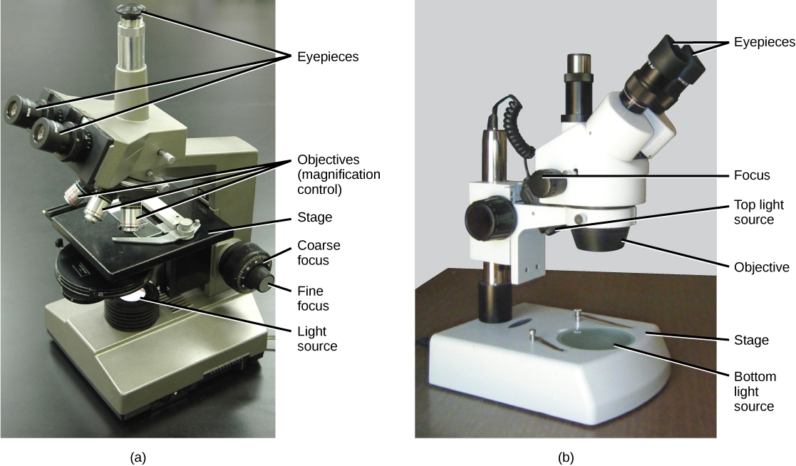
There are several different types of microscopes that can be used in a biology lab, each with its own strengths and limitations. The most common types of microscopes used in biology are light microscopes, electron microscopes, and fluorescence microscopes.

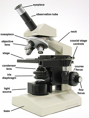
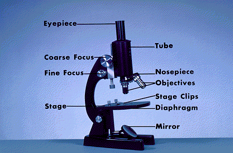
Light microscopes use visible light to magnify and illuminate the specimen, and are the most commonly used type of microscope in biology labs. They can be further divided into several subtypes, including compound microscopes, which use lenses to magnify the specimen, and stereo microscopes, which use two separate optical paths to produce a three-dimensional image.

Electron microscopes use a beam of electrons instead of light to magnify and illuminate the specimen. They can achieve much higher magnifications than light microscopes, but they also require special equipment and techniques to prepare the specimen for viewing.

Fluorescence microscopes use fluorescent dyes to label specific structures or molecules in the specimen, which can then be visualized using a special filter. This technique is useful for studying specific structures or processes within a cell or tissue.

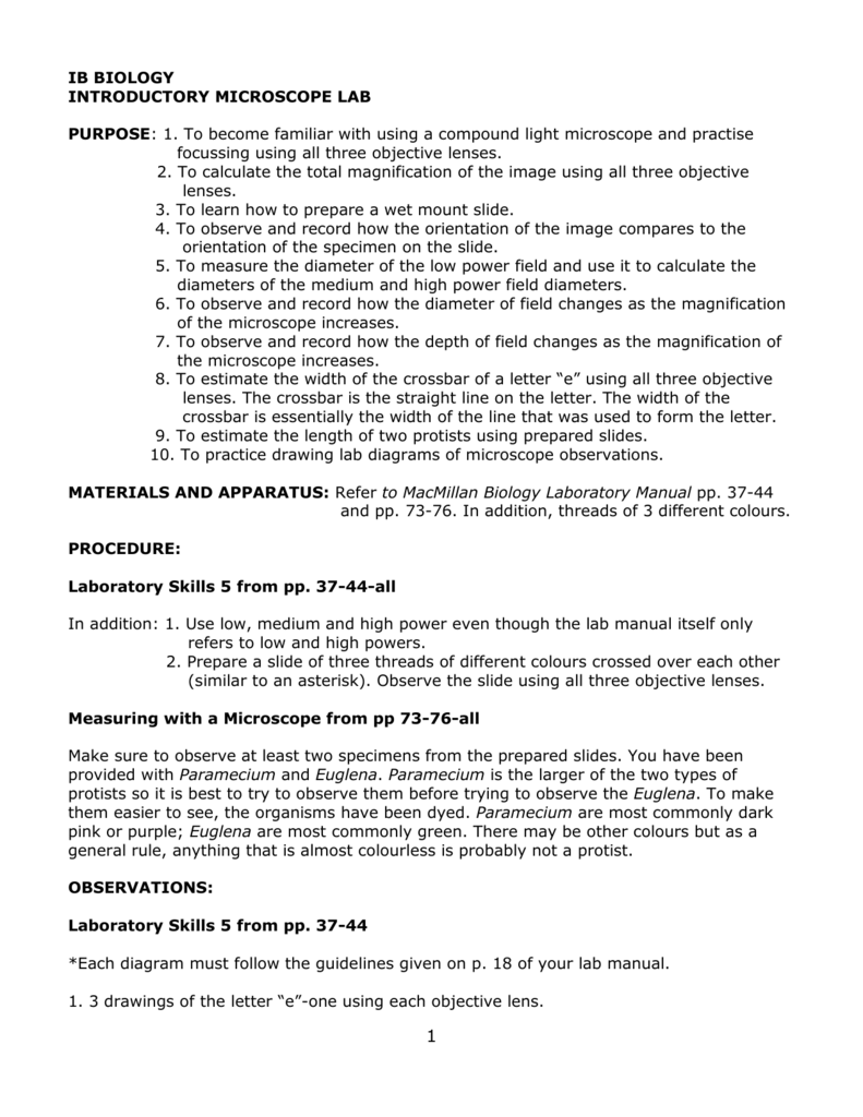
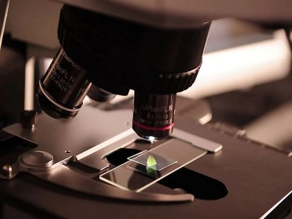
In a microscopy lab report, the student should first describe the equipment and techniques used to prepare and view the specimen. This should include details about the type of microscope used, the type of sample, and any special techniques or stains used to prepare the sample.

Next, the student should describe the results of the experiment, including any observations made about the specimen. This should include detailed descriptions of the structures or features observed, as well as any measurements or other data collected.

Finally, the student should discuss the implications of the results, including any conclusions that can be drawn based on the observations made. This may include discussing the importance of the structures or processes observed, or comparing the results to previous research on the same or similar topics.

Overall, a microscopy lab report is a detailed account of an experiment using microscopy to study small structures or organisms in biology. It should include a clear description of the equipment and techniques used, a thorough analysis of the results, and a discussion of the implications and significance of the observations made.

LAB REPORT 1 webapi.bu.edu

microscopy lab report biology

Do Not touch the coarse focus knob, instead use the fine focus to resolve the picture. The divisions of the two scales are not equal, as a result perfect overlapping of lines marking the divisions of the scales appear only at a few points. Add a drop of water. Look through the eye piece and turn the coarse adjustment for focusing. Cover the scope with the dust cover. In this lab, we are going to learn the proper use and handling of the microscope. As an example, the size of red blood cell is 6 to 8micrometer.

Next

Microscope

microscopy lab report biology

Show your formula and all work. Stage micrometer: It is a thick glass slide. Locate the diaphragm under the stage. If you view green material, it is probably some sort of plant. Now, move it to the lower left side of the stage. One of the main differences between primary and secondary journal articles is their source.

Next

Project Report on Microscope

microscopy lab report biology

Hypothesis The process known as wet-mount can be used to prepare a specimen on a slide which can be viewed with a compound light microscopes to produce an enlarged image. Remember with a compound light microscope you are magnifying with two lenses, so to calculate the total magnification you multiple the objective magnification by the ocular magnification. Cover the scope with the dust cover. Do the same for the high power objective. In artificial light the plain surface is used. If required the mirror can be taken out from the hole in the base of the microscope and a sub-stage lamp fitted. In this experiment, we used compound light microscope and stereomicroscope.

Next

Introduction to the Microscope Lab

microscopy lab report biology

It oper­ates in the way as in the coarse adjustment, but the movement of the body tube is only slight with the turning of the pinion. Only a minimal amount of adjustment is usually necessary. The adjustment has a large head. There are many times more microorganisms on Earth than all the other organisms put together. You are to follow the step-by-step sequence as your instructor indicates. The compound microscope, which is used for this experiment has two lenses, which will magnify. What is the size in micrometers of the microbe? Procedure Obtain a colored threads slide, and view it under scan or low power.

Next

Biology Research: Microbiology and Microscopy Lab Report, Lab Report Download Sample

microscopy lab report biology

The arrangement is to regulate the amount of light passing through the object. . Remove the slide and clean it up. It usually includes the sampling method, experimental design, analytical tools i. Move it and record the changes in light intensity as you do so. Indicate and describe a major way the stereomicroscope differs from the compound light microscope in terms of its use.

Next

BIOLOGY 101L: Laboratory Report 1: The Microscope Flashcards

microscopy lab report biology

. Microscope Lab: Introduction: Microscopes are used to enlarge a small object in order for it to be observed. Microorganisms are organisms that are too small to be seen with the human eye. Our mission is to provide an online platform to help students to share notes in Biology. Look to see if there is anything moving. Notice that the microscope has two light sources, one from the base and one from above. Turn on the light and open the diaphragm to produce a bright circle of light.

Next

Biology Lab Report

microscopy lab report biology

Microscope is an important tool for the scientist in many ways. To determine field of view. A microscope has a 20 X ocular eyepiece and two objectives of 10 X and 3 X respectively: a. Stage: It is a sort of squarish or rectangular platform with 10 to 1 2 cm sides and having a circular hole at the centre. The specimens are not always mounted on a slide.

Next

Biology Microscope Lab Report

microscopy lab report biology

Turn the nose piece to bring the low power objective lens in a line with the body tube. This microscope will produced 2-dimensional images Stereomicroscope or dissecting microscope is often used to study the surface of solid specimens or to carry out close work of dissection. Explain what the microscope user may have to do to combat the problems incurred in question 7. The objectives and eye pieces are changeable. Explain why the specimen must be centered in the field of view on low power before going to high power. Turn off the microscope and wind up the wire so it resembles its original position.

Next

biology .pdf

microscopy lab report biology

Describe the changes in the field of view and the amount of available light when going from low to high power using the compound microscope. It includes an overview of the methods, results, Discussion, and even the recommendation for its readers. Its primary purpose is to show whether the problem statement methods are credible enough. If the body tube is of standard length, i. We use microscopes to enlarge specimens for our investigation. The divisions of the micrometre are always smaller than those in the oculometre. The water should be sucked under the slide if done properly.

Next