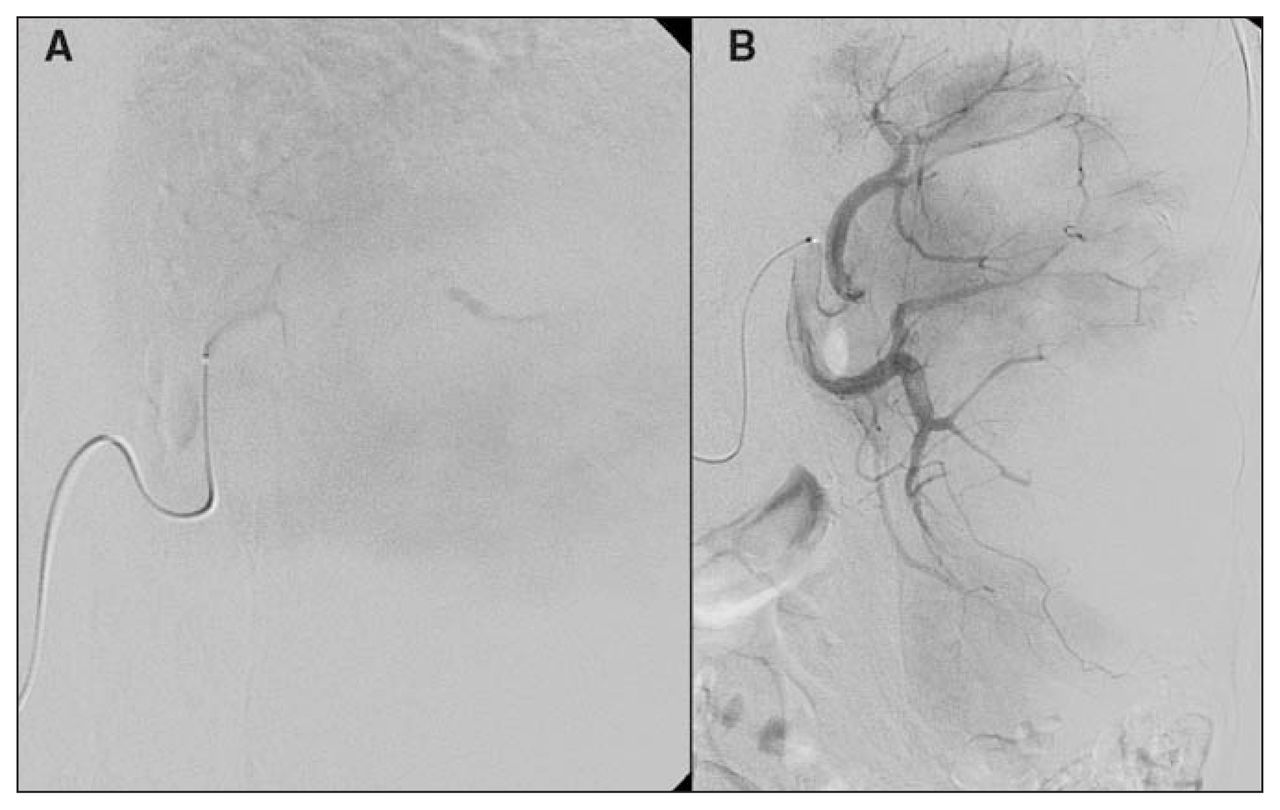
A mesenteric angiogram, also known as a mesenteric arteriogram, is a diagnostic procedure used to visualize the blood vessels in the mesentery, a thin layer of tissue that supports the intestines. The procedure is typically performed using X-ray imaging and an injection of contrast material, which allows the blood vessels to be seen more clearly on the images.
During a mesenteric angiogram, a small incision is made in the patient's groin, and a thin, flexible tube called a catheter is inserted into a blood vessel. The catheter is then guided through the blood vessels until it reaches the mesentery. Once in place, a contrast material is injected through the catheter and X-ray images are taken to visualize the blood vessels.
The procedure is typically performed to diagnose and evaluate conditions such as blockages or abnormalities in the mesenteric blood vessels, which can cause abdominal pain, abdominal swelling, and other symptoms. It may also be used to assess the blood flow to the intestines or to guide treatment such as the placement of stents to improve blood flow.
Mesenteric angiography is generally a safe procedure, but like any medical procedure, it carries some risks. These may include bleeding, infection, and allergic reactions to the contrast material. The patient may experience some discomfort during the procedure, but it is generally well tolerated.
In conclusion, a mesenteric angiogram is a valuable diagnostic tool for evaluating the blood vessels in the mesentery and can help guide treatment for a variety of conditions. It is a safe and effective procedure that has helped many patients receive the care they need.
Mesenteric angiography Information
Gated subtraction fast spin-echo: An imaging technique that subtracts two fast spin echo sequences acquired at systole and diastole. Flow dependent MRA can be divided into different categories: There is Time-of-flight TOF or inflow angiography, uses a short echo time and flow compensation to make flowing blood much brighter than stationary tissue. If the tumor blood vessels are visible, the artery infusion chemotherapy is selected. In some patients, the IVC is moderately compressed in its retrohepatic course by the enlarged liver and, in particular, by a hypertrophied caudate lobe Fig. It was decided to proceed with manual compression for hemostasis. The radiologist can then pinpoint and treat the source.
Mesenteric Angiogram

Superselective catheterization for intervention is usually accomplished with coaxial microcatheters. Retrieved 27 July 2021. Larry Jameson and Joseph Loscalzo. Certain treatments can be done during this procedure, including dissolving a blood clot with medicine, opening a partially blocked artery with a balloon, or placing a stent into an artery to help hold it open. An intravenous IV line will be started in your arm or hand and you will be connected to a monitor to record your heart rate, breathing, and blood pressure. Repeat the cycle at the same time for impact infusion chemotherapy. Orloff, in Blumgart's Surgery of the Liver, Pancreas and Biliary Tract Fifth Edition , 2012 Hepatic Angiography and Pressures The diagnostic study of greatest value in BCS, particularly if surgical therapy is contemplated and venous pressure measurements are required, is angiographic examination of the IVC and hepatic veins with pressure measurements Clain et al, 1967; Kreel et al, 1967; Redman, 1975; Tavill et al, 1975.
Magnetic resonance angiography

These include the development of flexible, hydrophilic, torque control guidewires and catheters that have low coefficients of friction, which allow them to follow wires around complex or tight curves more readily. Selective angiography superior mesenteric artery: Extraluminal calcification noted in the ostial and proximal portion the vessel luminal irregularities but no resultant stenosis noted TIMI-3 flow noted. It is moved into the artery, and up through the main vessels of the belly area until it is properly placed into a mesenteric artery. Superior Mesenteric Angiography The venous phase of superior mesenteric angiography demonstrates the characteristic distortion and abrupt diminution of second and third order portal venous radicles with occasional complete cut off the channels, also suggesting small venous occlusion. Material and Methods: This is an institutional review board-approved, retrospective study of 20 patients who underwent PMA between 2014 and 2019.