Maxilla anatomy. Maxillary Bones 2022-11-06
Maxilla anatomy
Rating:
4,2/10
1352
reviews
The maxilla, also known as the upper jawbone, is a paired bone that forms the upper part of the facial skeleton in humans and other mammals. It is located between the mandible, or lower jawbone, and the forehead. The maxilla is a complex bone that plays a vital role in several key functions, including supporting the teeth, shaping the face, and facilitating speech and swallowing.
One of the main features of the maxilla is its multiple projections, which help to give the face its unique shape. The most prominent projection is the maxillary prominence, which is located on the midline of the face and forms the upper part of the nose. The maxilla also has two zygomatic processes, which extend outward and contribute to the formation of the cheekbones.
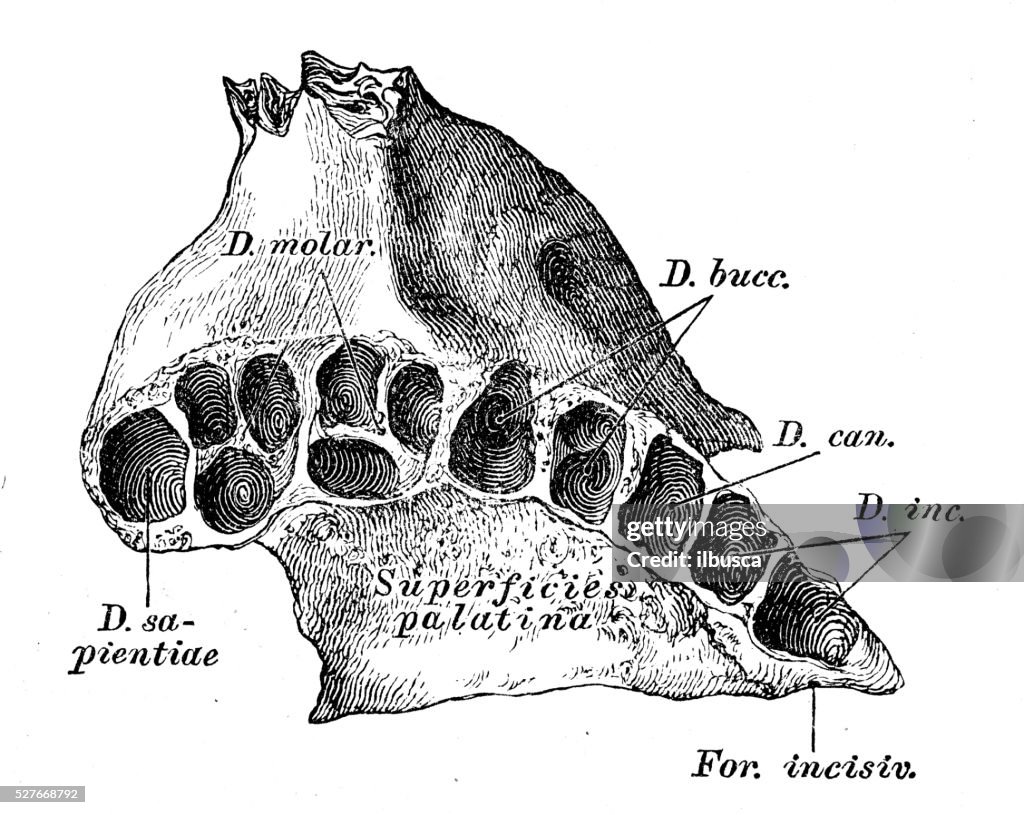
In addition to its role in shaping the face, the maxilla is also an important component of the dental arch. It contains sockets, or alveoli, that hold the upper teeth in place. The maxilla also has a central palatine process, which helps to form the roof of the mouth and separates the nasal cavity from the oral cavity.
Another important function of the maxilla is its role in speech and swallowing. The maxilla has several small bones called the palatine bones, which are connected to the tongue and help to shape the sounds of speech. The maxilla also has a soft palate, which helps to seal off the nasal cavity during swallowing to prevent food and liquids from entering the nasal passages.
Overall, the maxilla is a vital bone that plays a crucial role in several key functions, including shaping the face, supporting the teeth, and facilitating speech and swallowing. It is an intricate and complex bone that is essential to the proper functioning of the human body.
Maxilla Overview & Anatomy
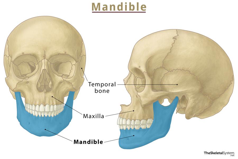
In newborns the maxilla is much longer horizontally than vertically, compared to adults. Allergic conditions can cause persistent inflammation and swelling of the sinuses and in some cases leads to abnormal growths inside of the sinus cavities called polyps which require surgical removal. We avoid using tertiary references. The stages of maxilla development are listed below. It is classified as a ''paired'' facial bone, meaning that two maxillae on either side of the face are fused in the middle. But the interradicular septa are bony ridges forming compartments in dental alveoli for the roots of the teeth in both the upper and lower jaw bones. Unlike the maxilla, the mandible has a joint and can move up, down, and side to side.
Next
Maxillary Bones

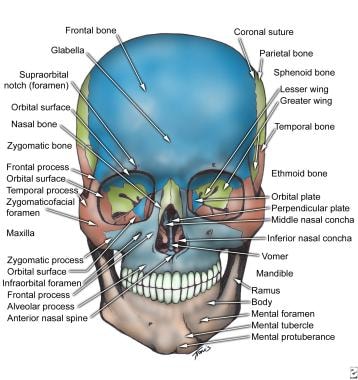
Maxilla The maxilla, also known as the upper jaw, is a vital This bone consists of five major parts, one being the body and four being projections named processes frontal, zygomatic, palatine, alveolar. Therefore, the maxilla has a variety of functions. Resetting the maxilla may require surgery. The masseteric artery accompanies the The pterygoid arteries are small branches that vary in number. It is similar to the mandible, the bone of the lower jaw, which hosts the lower teeth.
Next
Maxilla: Bone Anatomy, Function, and Surgery Procedures
This often happens due to injuries to the face, such as from falling, a car accident, getting punched, or running into an object. You may also need to have multiple surgeries depending on your injuries. Borley: Instant Anatomy, 4th edition, Wiley-Blackwell 2010 , p. Development of the maxilla begins around the 6th week of gestation and continues until an individual is between 14 and 15 years old. However, the remaining 10 branches, from the first and third parts, go through foramina in bones.
Next
Maxilla: Anatomy, function and clinical notes

Your doctor will determine when and how frequently they want to see you after surgery and once you are home. The roots of the teeth form grooves that extend up the anterior portion of the maxilla. Depending on your injuries, it may take two to four months or more. The alveolar yokes or juga alveolaria are eminences on the outer surfaces of the jaw bones produced by the projections of the dental alveoli. There are prominences in the bone where the tooth roots are.
Next
Maxilla
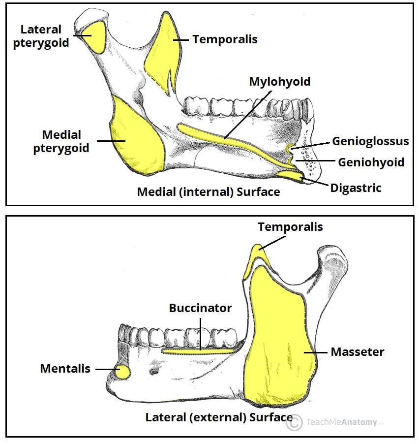
Find out how you can do just that by Extradural hematoma This is also called epidural haematoma, and it is the accumulation of blood in the epidural space due to injury, for example from a road traffic accident or sports injury, involving the middle meningeal artery. Since the maxilla has no joints and is immobile, the lower jaw does all the movement of mastication. The information we provide is grounded on academic literature and peer-reviewed research. Many facial muscles are connected to the maxilla on both its inner and outer surfaces. A Le Fort fracture is an example of a very serious maxilla fracture. It also articulates with the upper teeth. Her areas of interest include marine biology, ecology, genetics, and environmental science.
Next
Maxillary artery: Branches and anatomy

The inferior alveolar artery -  runs inferiorly and anteriorly towards the mandibular body of the mandible. It hosts the upper teeth which are essential for chewing. Depending on the extent of injury to your face, head, mouth, teeth, eyes, or nose, you may need a variety of specialists including, eye surgeons, oral surgeons, neurosurgeons, plastic surgeons, or ENT ear, nose, throat surgeons. The lower portion of the maxilla is connected to the upper teeth through the alveolar process. It arises from the same fetal tissue that forms the nose while the rest of the maxilla is formed by the fetal maxillary processes that form the rest of the bone.
Next
Maxilla: Anatomy, Function and Treatment

 It arises within the infraorbital canal where it descends to supply the The  pharyngeal artery supplies structures such as the nose. Because of the bone's location and proximity to important structures such as the airways, eyes, brain, and teeth, injuries to the maxilla can be serious. The extension to articulate with the zygomatic bone is known logically as the zygomatic process of the maxilla. Recovery could take between a few weeks to several months. Bones take a long time to heal. Posteriorly it forms the lacrimal groove together with the The zygomatic process of the maxilla grows laterally and meets the Lastly, the palatine process is a horizontal extension on the medial side of the bone constituting the roof of the mouth and the floor of the nasal cavity. Test yourself with our Learn the anatomy and function of the skull bones here: Bones of the skull Explore study unit Articulations The maxilla articulates with numerous bones: superiorly with the posteriorly with the medially with the inferior nasal concha and laterally with the zygomatic bone.
Next
Maxillary sinus: Anatomy and structure
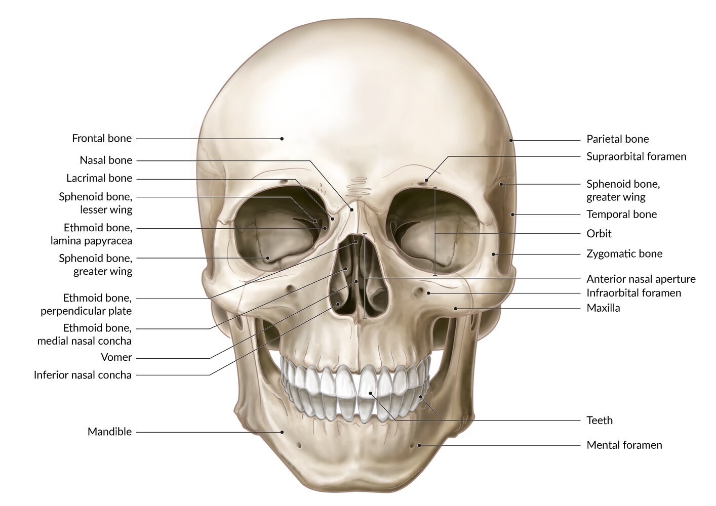
This sinus extends over the premolars and molars in the adult and superiorly to the base of the orbit. These form fossa between them. The body has several extensions referred to as processes. The jaw allows for chewing, the maxilla's primary function. The alveolar process is an inferior extension of the maxilla with a rather porous structure. This explains why cleft lips and clefts of the anterior palate are in the area of the canines rather than in the midline while clefts in the more posterior sections of the palate are in the midline.
Next
Maxilla Anatomy Flashcards
It continues its path by running deeply to the lower head and passes forward between the two heads of the lateral pterygoid muscle to break into its terminal branches at the pterygopalatine fossa. Here it gives off its posterior lateral nasal branches. Since her graduation in 2017, she has been involved in various ecological research projects in Michigan, Wisconsin, Minnesota, Alaska, and Massachusetts. It makes up a significant portion of the face and articulates with many other facial bones. This is actually a single opening at the confluence of the two incisive canals which are also referred to as the nasopalatine canals. Damage to the maxilla typically occurs from blunt force trauma to the face.
Next

In the 7th week of fetal life one differentiates between the maxilla and premaxilla or incisive bone. Maxilla fractures and other fractures that occur to the front of the face are also known as mid-face fractures. An individual who experiences a maxillary fracture will likely experience pain, swelling, and bruising. This is also useful since out of the 15 branches of the maxillary artery, the 5 branches from the second part part on the lateral pterygoid muscle are regarded as branches to soft tissues and they do not course through foramina in bones. Eventually, the maxilla is formed via ''pneumotization'', a simultaneous process of bone resorption and deposition. If you experience any trauma to your face or head, see your doctor right away.
Next