Main components of a light microscope. The Parts of a Light Microscope 2022-10-29

Main components of a light microscope Rating: 6,2/10 1242 reviews

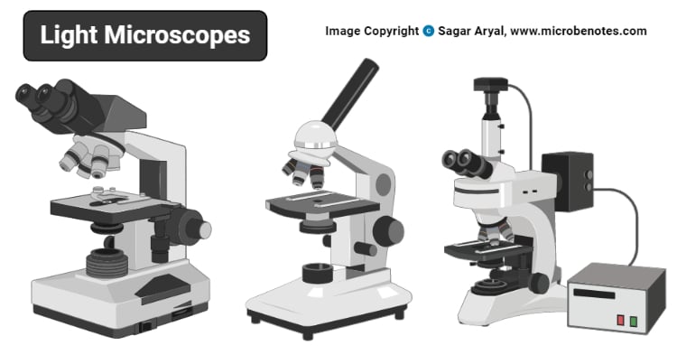
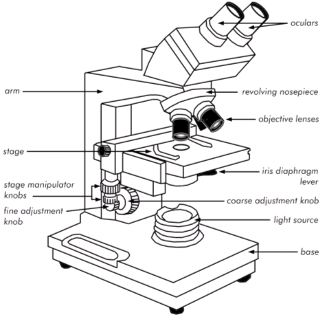
A light microscope is a scientific instrument used to magnify and examine small objects or structures that are not visible to the naked eye. It uses light as its source of illumination, and is an essential tool in many fields of science and medicine, including biology, physics, chemistry, and engineering. There are several main components of a light microscope, each of which plays a specific role in the functioning of the instrument.

The first component of a light microscope is the objective lens. This is a small, high-powered lens that is located on the end of the microscope near the specimen being viewed. The objective lens is responsible for gathering light from the specimen and focusing it onto the eyepiece lens. There are typically several objective lenses of different magnifications available on a microscope, allowing the user to select the best lens for the task at hand.

The second component of a light microscope is the eyepiece lens. This is the lens that the user looks through to view the specimen, and it is located at the top of the microscope. The eyepiece lens magnifies the image produced by the objective lens, allowing the user to see the specimen in greater detail. Most light microscopes have a single eyepiece lens, but some models have two eyepieces for stereo viewing.

The third component of a light microscope is the stage. This is the platform on which the specimen is placed, and it is located below the objective lens. The stage is typically equipped with a movable stage clamp or stage micrometer, which allows the user to accurately position the specimen under the objective lens. The stage may also have a built-in light source, such as a halogen lamp, to illuminate the specimen.

The fourth component of a light microscope is the condenser lens. This is a lens located below the stage that focuses light onto the specimen. The condenser lens helps to evenly illuminate the specimen and increase the contrast of the image. Some light microscopes have a diaphragm or iris located below the condenser lens, which allows the user to adjust the amount of light passing through the lens.

The fifth and final component of a light microscope is the base. This is the foundation of the microscope and provides support for the other components. The base is typically made of a sturdy material, such as metal or plastic, and may also include a mechanical stage for precise movement of the specimen.

In conclusion, a light microscope is a sophisticated instrument that allows scientists and researchers to view small objects and structures in great detail. It is composed of several main components, including the objective lens, eyepiece lens, stage, condenser lens, and base, each of which plays a vital role in the functioning of the microscope.

Components Of The Microscope Flashcards

main components of a light microscope

Microscopy works strictly with a factor of resolution whereby resolution is the ability of a lens to be able to differentiate small objects that are closely packed together. It captures light from an external source of a low voltage of about 100v. Fundamental to the understanding of image formation in the microscope is the action of individual lens elements that comprise the components in the optical train. Most light microscopes will come with either an iris diaphragm or a disc diaphragm. For mechanical stages, you can move the specimen on the X-Y axis using two knobs on the bottom-right of the stage. Figure 2: The angular aperture of a lens.

Next

What are the main components of a microscope?

main components of a light microscope

What are the 4 types of functions? This is a knob behind the focus knobs that stops the stage from being raised so high that it will bump against the objective lenses. Some high-quality microscopes have an articulated arm with more than one joint allowing more movement of the microscopic head for better viewing. Presented in Figure 1 is a typical microscope equipped with a trinocular head and 35-millimeter camera system for recording photomicrographs. In a fixed tube length microscope, this projection distance is about 160 millimeters. They are the most common type of microscope and are used in many fields, such as biology, medicine, and materials science.

Next

Microscope Optical Components

main components of a light microscope

Aspherical lenses ensure that no aberrations occur. Excitation wavelengths that manage to pass through the dichroic mirror are blocked by the barrier emission filter. Stage Adjustment Knobs Fine vs Coarse Focus Knobs The stage adjustment knobs allow you to raise and lower the stage to achieve focus. This is really useful for people who usually wear glasses or have different vision in each eye. For the objective and eyepiece just described, the total lateral magnification would be about 200x 10x eyepiece multiplied by the 20x objective. For a compound light microscope, there are usually two sets of ocular lenses in a purchase: 10X and 20X. All of the parts of a microscope work together — The light from the illuminator passes through the aperture, through the slide, and through the objective lens, where the image of the specimen is magnified.


Next

What Is Light Microscope and Its Types with Examples

main components of a light microscope

What are the magnifying parts of microscope? As it exits from Lens a , the plane wave is tilted with respect to the lens axis by an angle α. These lenses will retract if they accidentally tap against the specimen slide to protect the lenses from damage. Then the phase was somewhat lowered and the high power 40X aim lens was selected. Types of light microscopes optical microscope With the evolved field of Microbiology, the microscopes used to view specimens are both simple and compound light microscopes, all using lenses. The wavelength that excites the specimen and induces the fluorescence is not allowed to pass the filters placed between the objective lens and the eye.

Next

Light Microscope : Main Parts of Light Microscope

main components of a light microscope

You can push down on the back end of the stage clip to open it. All of the imaging components in the optical microscope are governed by the basic geometrical relationships described above. Most objectives are corrected to work within a narrow range of image distances, and many are designed to work only in specifically corrected optical systems with matching eyepieces. Figure: Diagram of parts of a microscope There are three structural parts of the microscope i. A real image can be visualized when projected onto a screen, captured on photographic film emulsion, or organized into a digital array by the photodiode elements of a charge-coupled device CCD.

Next

What are the three main parts of a microscope?

main components of a light microscope

The function of the coarse focus is to capture the exact distance roughly and quickly. Microscopes can be simple or complex in design, and some can do more than one type of microscopy, each of which reveals slightly different information. Head Siedentopf vs Sliding The head of a microscope is the housing that holds the ocular tubes and eyepieces. The angle a can be increased either by shortening the distance between the lens and the object or by increasing the diameter of the lens. This description defines the parts of a microscope and the functions they perform to enable the visualization of specimens.

Next

What are the parts of a light microscope and their functions?

main components of a light microscope

Wavetrains of light leaving the infinity-focused objective are collimated, allowing introduction of auxiliary components, such as differential interference contrast DIC prisms, polarizers, and epi-fluorescence illuminators, into the parallel optical path between the objective and the tube lens with only a minimal effect on focus and aberration corrections. It Is, therefore, a measure of how much of the illumination that leaves the specimen actually passes through the lens. Since, living cells are largely transparent, it is not always easy to see much internal structure when viewing even very large cells in the ordinary light microscope. Extensions of the refracted and incident ray intersect at a surface within the lens located at distance a from the source S 1. The main difference between light microscope and electron microscope is that beam of electrons is used for magnifying the image of an object while visible light is used in the light microscope to magnify images of tiny areas of materials or biological specimens. How to use a compound light microscope step by step? They play a major role in ensuring clear sharp images are produced with a high magnification of 400X and above.

Next

Light Microscope

main components of a light microscope

Eyepiece Lens: the lens at the top that you look through, usually 10x or 15x power. This gives contrast for easy visibility under the microscope. When observing the image through the eyepiece, the image-forming beam diverging out through the eyelens appears to originate from the virtual source located at Image Plane 3'. In terms of biology, it is one of the least invasive techniques for looking at living cells. From the source, light will pass through the specimen and into the objective lens, where a magnified image of the specimen will be formed. The larger the angular aperture, the more information the lens can transmit. The three components of light.


Next