In a frog embryo gastrulation. Gastrulation in Frogs 2022-11-07
In a frog embryo gastrulation Rating:
6,6/10
970
reviews
Gastrulation is a crucial process in the development of a frog embryo. It is the process by which the single-layered blastula transforms into a multi-layered structure called the gastrula. This process involves the movement and rearrangement of cells within the embryo, which ultimately determines the organization and differentiation of the various tissue types that make up the developing organism.
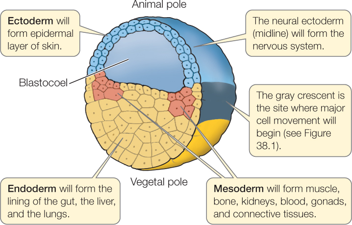
During gastrulation in a frog embryo, a series of cellular movements take place that result in the formation of three primary germ layers: the ectoderm, mesoderm, and endoderm. These germ layers will eventually give rise to all of the tissues and organs of the adult frog.
The ectoderm is the outermost germ layer and gives rise to the skin, nervous system, and sense organs. The mesoderm, which is located between the ectoderm and endoderm, gives rise to the skeletal, muscular, and circulatory systems. The endoderm, the innermost germ layer, gives rise to the lining of the digestive and respiratory systems.
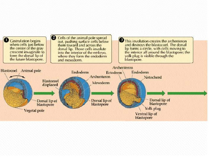
Gastrulation begins when a group of cells on the surface of the blastula called the primitive streak forms. The primitive streak is a linear indentation that runs along the length of the embryo. As cells at the tip of the primitive streak begin to migrate inward, they form a group of cells called the involuting cells. These cells move towards the center of the blastula and push the surrounding cells outward, forming the three germ layers.
The involuting cells that form the ectoderm do so by moving over the surface of the embryo, while the cells that form the mesoderm and endoderm migrate through the primitive streak. Once the germ layers have formed, they begin to differentiate into the various tissue types that will make up the developing frog.
Gastrulation is a complex and highly coordinated process that is essential for the proper development of a frog embryo. It is the first step in the formation of the various organs and systems that make up the adult frog, and it lays the foundation for all of the subsequent stages of development.
Gastrulation in Frogs
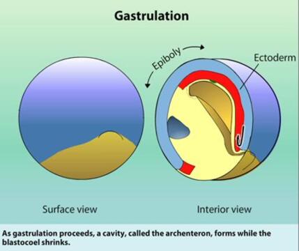
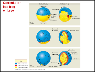
A deepening of the invagination results in a cavity called the archenteronor gastrocoel. ADVERTISEMENTS: On one side of the egg, in a position roughly corresponding to the marginal zone of the amphibian egg, there is a special type of cytoplasm; it does not contain much yolk, but it can be distinguished from the animal cytoplasm by its ability to be deeply stained by basic dyes. However during this process mesoderm and endoderm also undergo differentiation. The mesoderm gives rise to the notochord, which serves as a transient, organizing element for the developing embryo. The result of gastrulation is the formation of the three embryonic tissue layers, or germ layers.
Figure 1: The image above shows the process of transformation from a single-celled zygote to a gastrula. During this process the blastodermal cells begin to move. The opposite, dorsal, wall of the archenteron consists of cells of the marginal zone which have rolled into the interior over the dorsal lip of the blastopore. Different organisms have different types of amino acids. The outer layer of cells moves toward the blastopore, the location on the embryo where these cells invaginate to form the three embryonic layers, the ectoderm, the mesoderm, and the endoderm.
The embryo in this stage of development is called a gastrula. The term "Hox genes" applies to clusters of homeotic genes found in many different animals, including fruit flies. Immediately beneath the neural plate is a rod-shaped mesodermal structure called the notochord. By the end of cleavage, the embryo is called a blastula, which is a ball of cells surrounding a fluid-filled cavity called a blastocoel. From left to right.
Animal Development II: Gastrulation & Organogenesis
The mesoderm gives rise to the muscle cells and connective tissue in the body. This shift is an active process, an act of movement by the cells concerned. Possible responses to the signal may include synthesis of a particular protein or regulation of a particular enzyme. The histamine H1 receptor on target cells is a G protein-coupled receptor that activates phospholipase C in response to the binding of histamine. In fact, the upper limit of the marginal zone is none other than the limit to which the blastoderm is invaginated during the process of gastrulation. At the vegetal pole, it contains yolk filled with big cells. Drag the labels to their appropriate locations on the diagram.
The neural groovedeep-ens inside. This change in shape allows the G protein to bind to the H1 receptor, causing a GTP molecule to displace a GDP molecule and activating the G protein. In species in which there is a marked difference in color between a darkly pigmented animal hemisphere and a non-pigmented or lightly pigmented vegetal hemisphere, it can be noted that the light colored area is becoming smaller, and the dark-colored parts are reaching further down below the equator. During gastrulation, cell division slows dramatically, and cells are rearranged in a precise way, forming three germ layers. Organs are structures made up of two or more tissues organized to carry out a particular function, and groups of organs with related functions make up the different organ systems. School of Life Sciences.
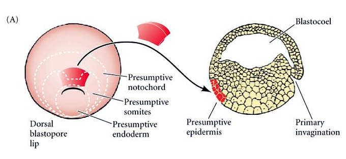
Involution enables the formation of the endoderm and mesoderm. During the gastrulation, the fate of the cells decides, and germ layers develop. The modified image is licensed under a CC BY-SA 3. The cells containing the basophilic cytoplasm are clearly discernible even as to shape. The mesodermal and endodermal cells gradually occupy their positions. Drag the labels to their appropriate locations on the diagram. As it does so, the presumptive notochord undergoes a very considerable elongation in the longitudinal direction and a corresponding contraction in the transverse direction.
The necessity of displacement of the parts so as to put them in the positions where they are situated in the adult animal is amply evident. For example, some of these proteins aid in elevating glucose levels in the blood, helping an animal to meet the demands of starvation or intense physical activity. The neural plate later thickens and it gets raised above the general level as ridges called neural folds. In the early stages of its formation this mass of cells is very loose and does not surround a cavity. These cells move to the interior. The external wall of the gastrula similarly takes part in the elongation of the embryo. Proteins produced in response to the cortisol signal function in the stress response.
Gastrulation in Gallus gallus Domestic Chicken. The two parts of the presumptive endoderm invaginate in different manners; the part lying in the marginal zone is mainly absorbed into the original pit-like invagination of the blastopore, and the vegetal region disappears from the surface when it becomes covered up by the contracting rim of the blastopore. The process of gastrulation in other frogs and toads appears to be intermediate between that found in urodeles and that of Xenopus. Gastrulation in Amphioxus: In Amphioxus, there are differences in the various regions of the egg cytoplasm which permitted Conklin 1932 to trace these regions into the later stages of development and thus to reconstruct a fate map, at least in rough outlines. This movement causes to create a dorsal lip. The information below was adapted from Development Step 3: Gastrulation At the end of gastrulation, in which the cells in the blastula rearrange themselves to form three layers of cells and form the body plan. Invagination Involution As invagination proceeds, there is the involution of marginal zone cells.
Gastrulation in Frog Embryo, Chick Embryo and Sea Urchin
The tops of the cells have microvilli that face the cavity from which substances will be absorbed. Dorsal view of human embryo. The cells of the presumptive notochord, which in the blastula lie in the inner marginal zone, accompany the endodermal cells in their forward thrust away from the rim of the blastopore and toward the animal pole but at no stage form part of the dorsal lining of the archenteron, and also at no stage are they on the exterior surface of the embryo. ADVERTISEMENTS: In this article we will discuss about the process of gastrulation in amphioxus and amphibians. This is a critical point in development because it is when the embryo transforms itself from a hollow sphere made from a single layer of cells into a multi-layered structure. Two types of signal receptors embedded in the cell's plasma membrane are G protein-coupled receptors and receptor tyrosine kinases.