The human abdomen is the area of the body between the thorax (chest) and pelvis. It is divided into four quadrants: the right upper quadrant, left upper quadrant, right lower quadrant, and left lower quadrant. These quadrants are further divided into nine regions: the epigastric, right hypochondriac, left hypochondriac, right lumbar, left lumbar, umbilical, hypogastric, right iliac, and left iliac.
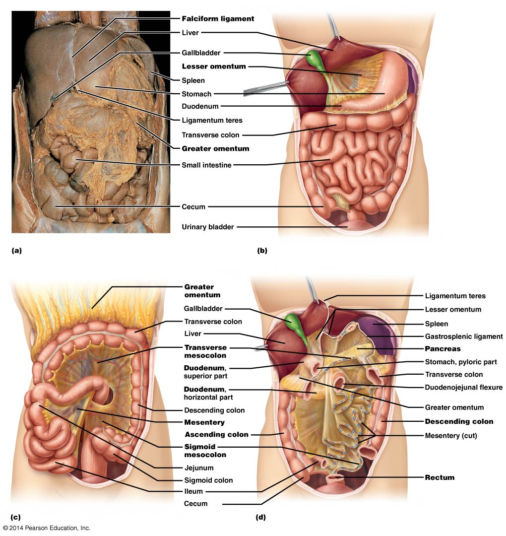
The organs located within the abdomen include the liver, stomach, small intestine, large intestine, pancreas, spleen, and kidneys. The abdominal aorta, which is the largest artery in the body, also runs through the abdomen.
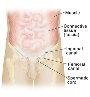
The abdomen is also home to several important muscles, including the rectus abdominis, which runs down the front of the abdomen and helps with movement and stability, and the external oblique muscles, which are located on the sides of the abdomen and help with rotation and lateral movement.
The abdomen is protected by the ribcage and vertebral column, which help to support the organs and muscles within it. It is also surrounded by a layer of fat, which helps to cushion and protect the organs.
Problems within the abdomen can often be diagnosed through physical examination and imaging techniques such as X-rays, CT scans, and MRIs. These techniques can help to identify issues such as organ damage or inflammation, as well as growths or masses.
Overall, the human abdomen is a complex and vital part of the body, housing a number of important organs and muscles that help to support many of the body's key functions. Understanding the anatomy and function of the abdomen is crucial for maintaining good health and identifying any potential problems that may arise.
Human Anatomy

Circle of sore in muscle, neck, stomach, throat and joint. The ureters are connected to the kidneys and are used to drain urine into the urinary bladder. Usually there is a single renal artery and vein, as well as a single ureter. Healthcare, sickness, disease concept, isolated on a white. The spleen functions to remove old and senescent red blood cells from circulation and acts as a secondary lymphoid organ, but does not aid in digestion. Although they are not large organs, the kidneys receive 25% of the cardiac output of blood, a testament to their critical function in maintaining appropriate blood volume, pressure, and concentration. The small intestine occupies the majority of the space of the abdominal cavity.
Human Abdomen Anatomy

Despite its misleading name, the large intestine is shorter about five feet than the small intestine, but it is larger in girth. Structures of the posterior abdomen include the kidney and ureters as well as some muscles that move the lower limb. Within the head of the pancreas, the common bile duct and the pancreatic duct join together briefly and empty into the duodenum through a common opening. In particular, the kidneys function to filter the blood of waste products, regulate blood pressure, and control the blood pH. Pyramidalis Muscle The pyramidalis muscle is a small, triangular-shaped muscle situated in front of the rectus abdominis in the lower portion of the abdomen. The transverse abdominus muscle and internal obliques affect posture by providing spinal support during rotation and lateral flexion, and stabilize the spine when standing.
Human Anatomy Abdomen Illustrations, Royalty

Abdominal surgery can be used to treat a number of conditions, including infections, tumours, inflammatory bowel disease or obstructions. Because this foramen is within the central tendon, it will get larger when the diaphragm contracts, because the central tendon is pulled taut. Normal capacity of the stomach is about one liter, though it can purportedly be stretched to a whopping four liters of capacity! When fat reaches the small intestine, the gall bladder is stimulated to release bile. Diet food, nutritions - protein, fat, carbohydrate, fit body vector illustrations. Medical emergency, unhappy and elderly guy in retirement in bedroom with abdomen illness, indigestion or constipation. They also secrete corticosteroids and androgens, among other hormones. The kidneys also help regulate levels of electrolytes, like salt and potassium, and produce certain hormones that play various roles throughout the body.
(204).jpg)


