External structure of human heart. Essay on Human Heart: Location, Structure and Other Details (with diagram) 2022-10-16

External structure of human heart Rating: 4,3/10 1648 reviews

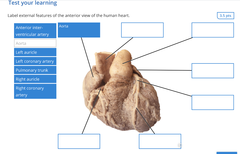
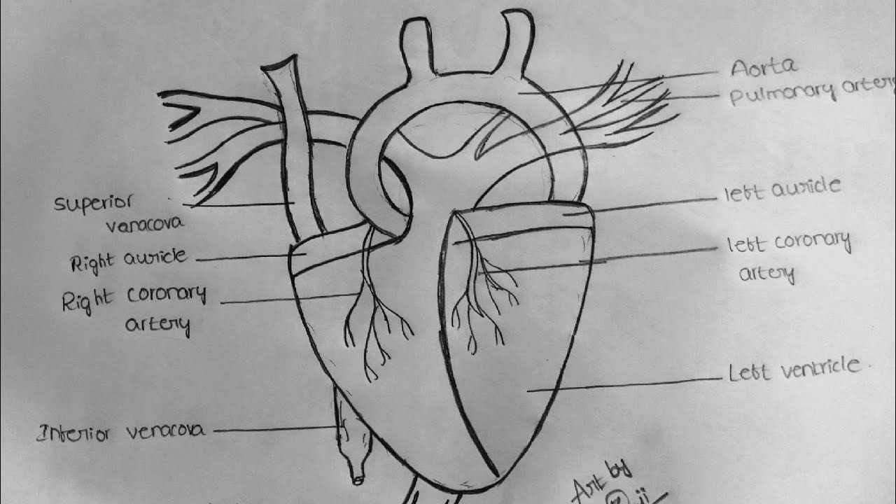
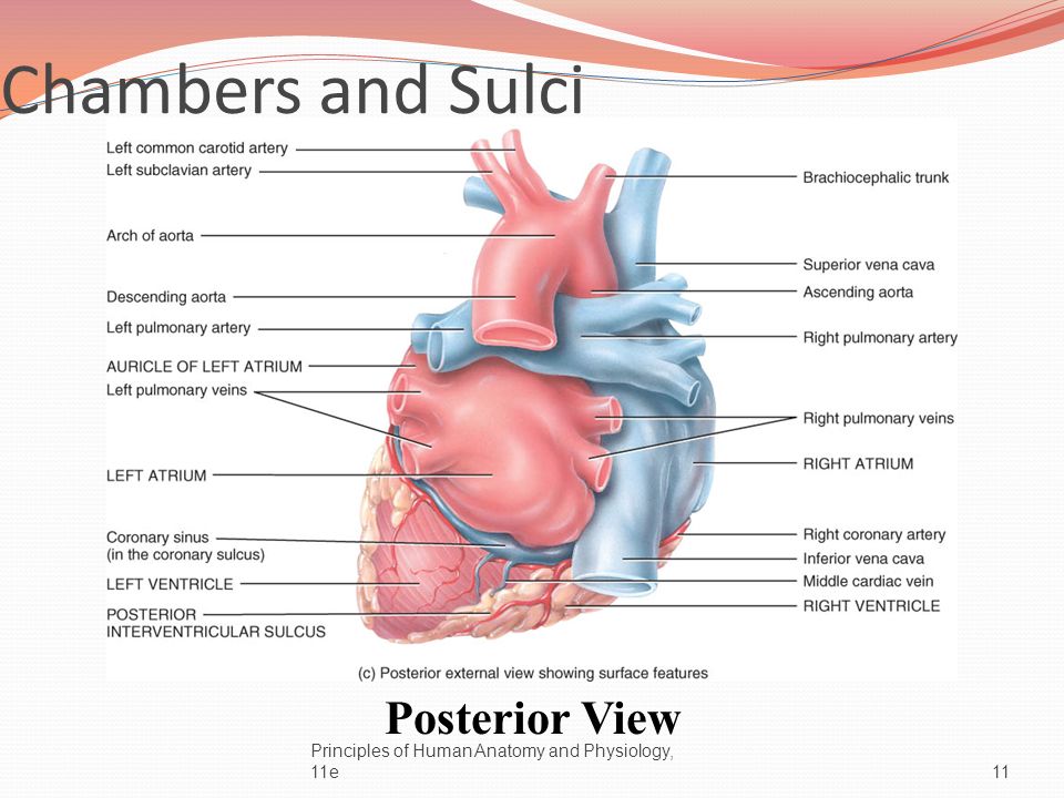
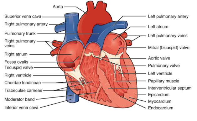
The human heart is a complex and vital organ that is responsible for pumping blood throughout the body. It is located in the thoracic cavity, between the lungs, and is protected by the ribcage. The heart is divided into four chambers: the left and right atria, and the left and right ventricles. The atria are the upper chambers of the heart, while the ventricles are the lower chambers.

The heart has a unique external structure that helps it to function effectively. The outer layer of the heart is known as the epicardium, which is a thin layer of tissue that covers the surface of the heart. The middle layer of the heart is known as the myocardium, which is a thick layer of muscle tissue that is responsible for contracting and relaxing to pump blood through the heart. The inner layer of the heart is known as the endocardium, which is a thin layer of tissue that lines the inside of the heart and helps to prevent damage to the heart muscle.

The heart is also surrounded by a network of blood vessels, including the aorta, the vena cava, and the pulmonary artery and vein. These blood vessels transport oxygenated and deoxygenated blood to and from the heart, respectively.

The heart has a unique system of electrical conduction that helps to coordinate the contraction of the heart muscle. This system includes the sinoatrial (SA) node, the atrioventricular (AV) node, and the Purkinje fibers. The SA node is located in the right atrium and acts as the heart's natural pacemaker, sending electrical impulses that stimulate the contraction of the heart muscle. The AV node is located in the interatrial septum and helps to coordinate the contraction of the atria and ventricles. The Purkinje fibers are located in the ventricles and help to transmit the electrical impulses throughout the heart muscle.

In summary, the human heart has a complex external structure that is essential for its function. It is made up of three layers of tissue, is surrounded by a network of blood vessels, and has a unique system of electrical conduction. All of these components work together to ensure that the heart is able to pump blood efficiently and effectively throughout the body.

The human heart (External and internal structure)

external structure of human heart

A stroke is a condition caused by the blockage of the flow of blood to the brain. The coronary arteries supply oxygenated blood to the heart, and the cardiac veins drain the blood once it has been deoxygenated by the tissues of the heart. Subclavian arteries : Supply blood to the arms. Papillary muscles act as anchors for chordae tendinae. Handbook of Cardiac Anatomy, Physiology, and Devices. What is the interior of heart? Pulmonary arteries carry deoxygenated blood to the lungs.

Next

Anatomy of a Human Heart

external structure of human heart

Sinus of valsalva: When the semilunar valves open towards aorta, a gap remains between the flaps and the wall of aorta which is called sinus of valsalva. Small veins are called venules. The waves of contraction is initiated by Sino-auricular node S. Also Refer:Â The Function of Heart The function of the heart in any organism is to maintain a constant flow of blood throughout the body. State of the Art Surgical Coronary Revascularization. Abdominal aorta is the largest artery.

Next

The external structure of human heart

external structure of human heart

Exploring in Yahoo I eventually stumbled upon this website. The heart is one of the essential organs in every organism. Definition of Heart: The heart is the essential organ in the cardiovascular system that is used for the pumping of the oxygenated and deoxygenated blood throughout the body. Other abnormalities are coronary arteries originating from a not typical sinus and then traveling either between the Aorta and pulmonary artery, or intramural. Atrio-ventricular valves: Auricles open into the ventricles through auriculo-ventricular apertures or AV apertures guarded by one way Atrioventricular valves or parachute valves and are located dorsally or posteriorly. The right fibrous trigone is the strongest part of the skeleton. Thus, The heart consists of four chambers, The right and left atria and the right and left ventricles.

Next

34

external structure of human heart

The outer layer is non distensible fibrous pericardium and inner layer is thin serous pericardium which further consists of outer parietal layer attached to fibrous pericardium and inner visceral layer adhered to the heart and is called epicardium. There is a fluid between the heart and the pericardial fluid. The ventricles are the chambers that pump blood and the atrium are the chambers that receive blood. Below are a collection of different diseases, to name but a few. The heart wall consists of 3 layers: Epicardium: thin external layer formed by visceral layer of serous pericardium. The walls of veins are thin, less muscular and non- elastic.

Next

Heart anatomy: Structure, valves, coronary vessels

external structure of human heart

In infants it is felt in the 3 rd intercostal space just lateral to the midclavicular line. Blood delivers oxygen and nutrients to every cell and removes the carbon dioxide and other waste products made by those cells. Node situated on inner wall of right auricle. External Structure of the human heart The heart is concaved shaped, dark red in color which is enclosed by the double-layered membrane called the pericardium. When the heart rate is faster than it starts to pump a higher amount of blood which makes the body ready for action during frightening with the supply of a higher amount of oxygen into the body.


Next

Anatomy of the human heart

external structure of human heart

Ade Owner of Ishwaranand Website. This may be associated with the reversal of all the abdominal organs, a clinical condition known as situs inversus. When ventricle contract and A. Similarly, the rate of the heart differs according to the situation. It also may include the major vessels immediately entering and exiting the heart, such as the aorta,….

Next

Internal and external structure of the Human Heart

external structure of human heart

It is lubricating fluid, which reduces friction between the pericardium and heart. Artery has thick, muscular and elastic walls. So on average, the weight of the heart is supposed to be 250-350 grams in adult males and 200-250 grams in adult females. ECG is measured when the heart activity is imbalanced or when it does not perform the rhythmic contraction and relaxation. Bundle of HIS: Starts from A. Aorta also has semilunar valves to prevent back flow into the left ventricle.


Next

What is internal and external structure of heart?

external structure of human heart

Dorsal aorta : it curls backwards and following arteries are given off: a H epatic artery : Supplies blood to the liver. Sometimes the pacemaker becomes faulty slow heart beat causing heart trouble. Exterior Chambers of the Heart. All content published on Kenhub is reviewed by medical and anatomy experts. External Structure : The heart Fig. When the left atrium contracts the blood enters the left ventricle through bicupsid valve. Thus, any defect in the valve to open and close may lead to blocking of the blood flow.

Next