Dark and bright field microscopy. Dark Field Microscopy: What it is And How it Works » Microscope Club 2022-10-26

Dark and bright field microscopy Rating: 5,2/10 464 reviews

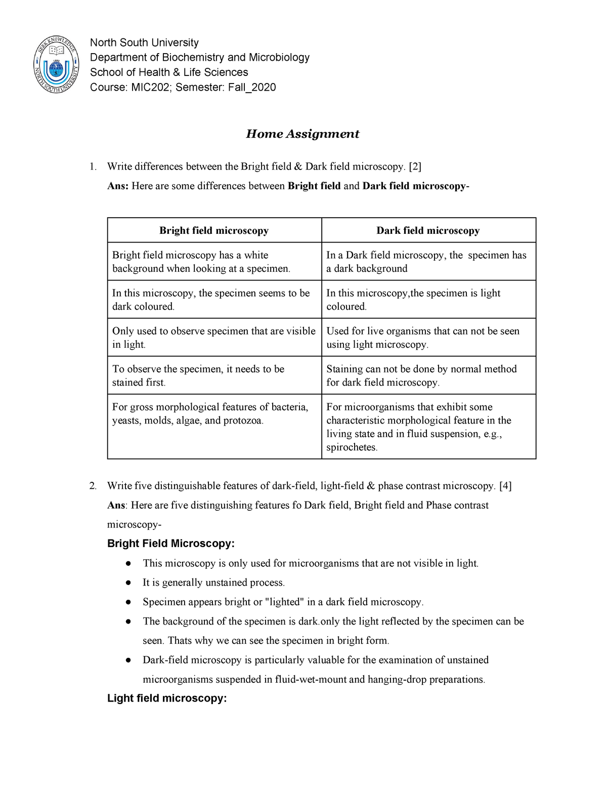
Dark field microscopy and bright field microscopy are two common techniques used in microscopy to visualize samples and obtain detailed images of their structures. Both techniques use light to illuminate the sample, but they differ in how the light is collected and analyzed to form the image.

Dark field microscopy is a technique that utilizes oblique illumination to visualize samples. In this technique, the light source is positioned at an angle to the microscope objective, so that the light is not directly transmitted through the sample. Instead, the light is scattered by the sample, and the scattered light is collected by the objective and focused onto the detector. This results in an image that appears dark, with the sample appearing as bright structures against a dark background.

Dark field microscopy is particularly useful for visualizing transparent or transparent samples, as well as for observing small particles or structures that may be difficult to see under bright field conditions. It is also useful for identifying the shape, size, and arrangement of cells and other structures within a sample.

Bright field microscopy, on the other hand, is a technique that uses direct illumination to visualize samples. In this technique, the light source is positioned directly above the sample, and the light is transmitted through the sample and the objective, and then focused onto the detector. This results in an image that appears bright, with the sample appearing as dark structures against a bright background.

Bright field microscopy is the most common technique used in microscopy, and it is particularly useful for visualizing samples that are opaque or have a high contrast with the surrounding medium. It is also useful for identifying the overall structure and organization of a sample.

In conclusion, both dark field microscopy and bright field microscopy are important techniques in microscopy, and they are used to visualize and study the structure and organization of samples at the microscopic level. Each technique has its own strengths and limitations, and the choice of which technique to use depends on the specific properties of the sample and the information that is desired.

Dark

dark and bright field microscopy

Darkfield Microscopy Darkfield microscopy is used in both light and electron microscopy. However, as staining is a must condition here, the proper process of staining should be known by the users. From a scientific standpoint, this is a very powerful technique. You can easily modify or adjust the settings of your microscope to illuminate the specimens under a dark field. ALSO READ: Overview Dark field microscopy is a simple yet useful and effective type of microscopy technique that illuminates the specimen in such a way that the background is dark and the specimen is well lit, thus making for a high contrast and high resolution image. A darkfield microscope offers a way to view the structures of many types of biological specimens in greater contrast without the need of stains.

Next

Dark Field Microscopy: What it is And How it Works » Microscope Club

dark and bright field microscopy

The specimens used are prepared initially by staining to introduce color for easy contracting characterization. It should also be placed closer to the condenser at low focusing power, and closer to the light source at high focusing power. This allows these faint rays to enter the objective. What is the disadvantage of dark field microscopy? In contrast, the portion of the ring-shaped light that passes directly through the specimen non-deviated or is reflected by the sample without scattering will not be collected by the objective. The low light levels observed in the finished image are, however, a drawback of darkfield. Both types of microscopes have their own advantages and disadvantages.

Next

What is bright field and dark field microscopy?

dark and bright field microscopy

Opaque disc The opaque disc is not present in it. What is difference between SEM and TEM? Lister the father of Joseph Lister invented the darkfield microscope, in which the standard brightfield Abbe condenser is replaced with a single or double-reflecting dark field condenser. This dark background provides a high degree of contrast and can make samples with difficult backgrounds stand out with relatively little effort. What is the difference between bright field and dark field microscope? Diffraction increases and the edges of the specimen becomes darker even though they are not in reality. This type of microscope contains a special condenser that scatters light and causes it to reflect off the specimen at an angle. They lack contrast with the surrounding medium. Suitability for amateur microscopy: High.

Next

Bright Field vs. Dark Field

dark and bright field microscopy

They also hold the microscope base which is the stand of the microscope. The result is a bright specimen on a black background. Dark field microscopes are designed to overcome these limitations. Yet, switching condensers based on the illumination type is impractical for everyday microscope use. The filter blocks the direct light of the microscope.

Next

What Is Darkfield Microscopy?

dark and bright field microscopy

Image source: Created with BioRender. Especially some crystallized structures are capable of that. Cell structures that appear transparent with brightfield illumination can be viewed with better contrast and detail using darkfield. This produces an enlarged clear image that is then magnified again by the eyepiece to form the primary image that is seen by the eyes. It can be used to look at blood cells and parts of a cell, tissue sections, yeast, bacteria, algae, various kinds of invertebrates, protists and metazoans, pond water, soil infusions, hay, precious stones such as diamonds, and fractures on metals.

Next

Brightfield Microscope (Compound Light Microscope)

dark and bright field microscopy

What are bright field images? There are, however, some limitations. Stained, fixed and live specimens are observed under a bright field microscope. Here the sample must be bright and the background of the sample need to remain dark. To brighten the digital image on the monitor, camera parameters such as exposure time and gain can be increased. Because most biological samples have little contrast, the image can appear bleached.

Next

Darkfield Microscopes

dark and bright field microscopy

However, there are some limitations to this type of microscope. Tiny pieces of fragmented wood take on an unusually beautiful appearance when illuminated under darkfield conditions with a transmitted light microscope. It works excellently on highlighting the details of smooth surfaces, minimally refractive specimens, and certain areas of the specimen that are usually obscured by shadows when viewed through a bright field microscope. Who invented dark field microscope? It depends on what you want to observe. Usability Using this microscope is quite easy. This technique is highly recommended due to its ease of use, low cost and strong visual effect. When to use bright field microscopy Bright field microscopy is best suited to viewing stained or naturally pigmented specimens such as stained prepared slides of tissue sections or living photosynthetic organisms.

Next

Differences Between Bright and Dark Field Microscopes

dark and bright field microscopy

Dark-field microscopy is a technique that can be used for the observation of living, unstained cells and microorganisms. Brightfield Microscope Definition It is the standard microscope that is used in Biology, Cellular Biology, and Microbiological Laboratory studies. Here the specimen is not needed to be stained. In general, objects imaged under the proper conditions of darkfield illumination are spectacular to see. By understanding these differences, you can choose the right microscope for your needs. All other light that passes through the specimen will miss the objective, thus the specimen is brightly illuminated on a dark background.

Next